Konstantinos C Soultanis, Konstantinos Tsiavos, Theodoros B Grivas, Nikolaos A Stavropoulos, Vasileios I Sakellariou, Andreas F Mavrogenis, Panayiotis J Papagelopoulos
{"title":"Reliability study for the Rib Index in chest radiographs of a control group.","authors":"Konstantinos C Soultanis, Konstantinos Tsiavos, Theodoros B Grivas, Nikolaos A Stavropoulos, Vasileios I Sakellariou, Andreas F Mavrogenis, Panayiotis J Papagelopoulos","doi":"10.1186/1748-7161-10-S2-S9","DOIUrl":null,"url":null,"abstract":"<p><strong>Background: </strong>The Rib Index, (RI), extracted from the double rib contour sign (DRCS) on lateral spinal radiographs to evaluate rib hump deformity, (RHD), in idiopathic scoliosis, (IS), patients, has been previously introduced. Although various papers using the RI have been published, no study on its reproducibility has been reported. The aim of this report is to estimate the variations of the RI in a number of a pair set of lateral chest radiographs (LCRs). The hypothesis was that the RI should have minimal variability for each subject having successive LCRs.</p><p><strong>Methods: </strong>Seventy randomized patients who were treated in the hospital for lung diseases (mainly pneumonia or other communicable lung diseases), were initially included in the study. Each of these patients had two successive LCRs (named A and B group of radiographs) at the radiological department of the hospital, by the same technician, during the course of their treatment. The radiation source - patient distance was constant. LCRs obtained at an incorrect patient's position, or from patients who underwent a thoracic intervention and all LCRs with symmetric hemi-thoraces were excluded from the study. The LCRs of 49 patients were deemed suitable for inclusion in the study. The RI was calculated in both (A and B) LCRs of each patient. The statistical analysis included the following techniques: paired t-test, Pearson correlation coefficient and intra- and inter-observer error using the formula (SD/√2)/2, where SD is this of the differences of the two sets of measurement (As-Bs). The SPSS v16 statistical package was used.</p><p><strong>Results: </strong>In the 49 pairs of LCRs there was no statistical difference of the RI, (paired t-test p< 0.314). The RI in the A and B group of LCRs was perfectly correlated (correlation coefficient = 0,924, p < 0.0001). The intra-observer error was 0.0080 while the inter-observer error 0.0213 in terms of 95% CI.</p><p><strong>Conclusion: </strong>The RI proves to be a reliable method to evaluate the thoracic deformity and the effect of surgical or non-operative treatment on the IS RHD. RI is a simple method, a safe reproducible way to assess the RHD based on lateral radiographs, without the need for any further special radiographs and exposure to additional radiation.</p>","PeriodicalId":21722,"journal":{"name":"Scoliosis","volume":"10 Suppl 2","pages":"S9"},"PeriodicalIF":0.0000,"publicationDate":"2015-02-11","publicationTypes":"Journal Article","fieldsOfStudy":null,"isOpenAccess":false,"openAccessPdf":"https://www.ncbi.nlm.nih.gov/pmc/articles/PMC4331769/pdf/","citationCount":"0","resultStr":null,"platform":"Semanticscholar","paperid":null,"PeriodicalName":"Scoliosis","FirstCategoryId":"1085","ListUrlMain":"https://doi.org/10.1186/1748-7161-10-S2-S9","RegionNum":0,"RegionCategory":null,"ArticlePicture":[],"TitleCN":null,"AbstractTextCN":null,"PMCID":null,"EPubDate":"2015/1/1 0:00:00","PubModel":"eCollection","JCR":"","JCRName":"","Score":null,"Total":0}
引用次数: 0
Abstract
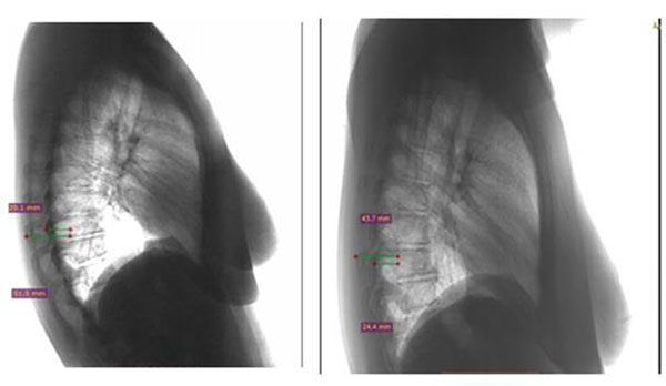
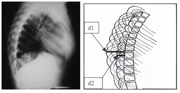
Background: The Rib Index, (RI), extracted from the double rib contour sign (DRCS) on lateral spinal radiographs to evaluate rib hump deformity, (RHD), in idiopathic scoliosis, (IS), patients, has been previously introduced. Although various papers using the RI have been published, no study on its reproducibility has been reported. The aim of this report is to estimate the variations of the RI in a number of a pair set of lateral chest radiographs (LCRs). The hypothesis was that the RI should have minimal variability for each subject having successive LCRs.
Methods: Seventy randomized patients who were treated in the hospital for lung diseases (mainly pneumonia or other communicable lung diseases), were initially included in the study. Each of these patients had two successive LCRs (named A and B group of radiographs) at the radiological department of the hospital, by the same technician, during the course of their treatment. The radiation source - patient distance was constant. LCRs obtained at an incorrect patient's position, or from patients who underwent a thoracic intervention and all LCRs with symmetric hemi-thoraces were excluded from the study. The LCRs of 49 patients were deemed suitable for inclusion in the study. The RI was calculated in both (A and B) LCRs of each patient. The statistical analysis included the following techniques: paired t-test, Pearson correlation coefficient and intra- and inter-observer error using the formula (SD/√2)/2, where SD is this of the differences of the two sets of measurement (As-Bs). The SPSS v16 statistical package was used.
Results: In the 49 pairs of LCRs there was no statistical difference of the RI, (paired t-test p< 0.314). The RI in the A and B group of LCRs was perfectly correlated (correlation coefficient = 0,924, p < 0.0001). The intra-observer error was 0.0080 while the inter-observer error 0.0213 in terms of 95% CI.
Conclusion: The RI proves to be a reliable method to evaluate the thoracic deformity and the effect of surgical or non-operative treatment on the IS RHD. RI is a simple method, a safe reproducible way to assess the RHD based on lateral radiographs, without the need for any further special radiographs and exposure to additional radiation.

 求助内容:
求助内容: 应助结果提醒方式:


