{"title":"Measurement of axial vertebral rotation using three-dimensional ultrasound images.","authors":"Quang N Vo, Edmond Hm Lou, Lawrence H Le","doi":"10.1186/1748-7161-10-S2-S7","DOIUrl":null,"url":null,"abstract":"<p><strong>Background: </strong>Axial vertebral rotation (AVR) is one of the important parameters to evaluate the severity and predict the progression of scoliosis. However, the AVR measurements on radiographs may underestimate its actual value. This pilot study investigated a new three-dimensional (3D) ultrasound method to measure AVR.</p><p><strong>Methods: </strong>Three cadaveric vertebrae T7, L1, and L3 were scanned with a 3D medical ultrasound system. Nine sets of ultrasound data, the vertebral rotation from 0 to 40° with 5° increments, were recorded from each vertebra. An in-house program was developed to reconstruct and measure the 3D vertebral images. The rotation of each reconstructed vertebra was determined by the angle between the line going through the centres of either laminae (L-L) or transverse processes (TP-TP) and a reference vertical plane. Three raters measured the rotation in 3 sessions, in which they used the mouse pointer to select the L-L or TP-TP according to their knowledge of vertebral anatomy. The program detected the 3D coordinates of these points and calculated the AVR. The intra-class correlation coefficients (ICCs) were used to calculate the intra-reliability and inter-reliability. The mean absolute difference (MAD±SD) and the range of difference (RD) between the actual values and the average measurements of each rater were computed to evaluate the accuracy of methods.</p><p><strong>Results: </strong>When rotation was greater than 30° for both L1 and L3, all raters found it difficult to determine one of the lamina areas due to the lack of ultrasound information in an area behind the spinous process. Therefore, the corresponding measurements were excluded. The ICC values of the intra-reliability (L-L, TP-TP) for the three raters were (0.987, 0.991), (0.989, 0.998) and (0.997, 1.000), respectively; meanwhile, the inter-reliability were 0.991 for (L-L) and 0.992 for (TP-TP). All ICC values were greater than 0.98 indicating both methods were highly reliable. The MAD±SD values (L-L, TP-TP) for the three raters were (1.5±0.3°, 1.2±0.2°), (1.6±0.3°, 1.3±0.3°), and (1.7±0.5°, 0.9±0.2°), respectively. The RD (L-L, TP-TP) were (0-4.5°, 0-3.5°), (0-5.1°, 0-4.3°), and (0-5.1°, 0-2.8°) for the three raters, respectively.</p><p><strong>Conclusions: </strong>The (L-L) and (TP-TP) methods could be used to measure AVR reliability from the 3D ultrasound images.</p>","PeriodicalId":21722,"journal":{"name":"Scoliosis","volume":"10 Suppl 2","pages":"S7"},"PeriodicalIF":0.0000,"publicationDate":"2015-02-11","publicationTypes":"Journal Article","fieldsOfStudy":null,"isOpenAccess":false,"openAccessPdf":"https://www.ncbi.nlm.nih.gov/pmc/articles/PMC4331767/pdf/","citationCount":"0","resultStr":null,"platform":"Semanticscholar","paperid":null,"PeriodicalName":"Scoliosis","FirstCategoryId":"1085","ListUrlMain":"https://doi.org/10.1186/1748-7161-10-S2-S7","RegionNum":0,"RegionCategory":null,"ArticlePicture":[],"TitleCN":null,"AbstractTextCN":null,"PMCID":null,"EPubDate":"2015/1/1 0:00:00","PubModel":"eCollection","JCR":"","JCRName":"","Score":null,"Total":0}
引用次数: 0
Abstract
Background: Axial vertebral rotation (AVR) is one of the important parameters to evaluate the severity and predict the progression of scoliosis. However, the AVR measurements on radiographs may underestimate its actual value. This pilot study investigated a new three-dimensional (3D) ultrasound method to measure AVR.
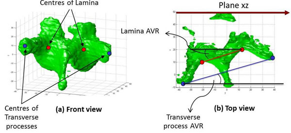
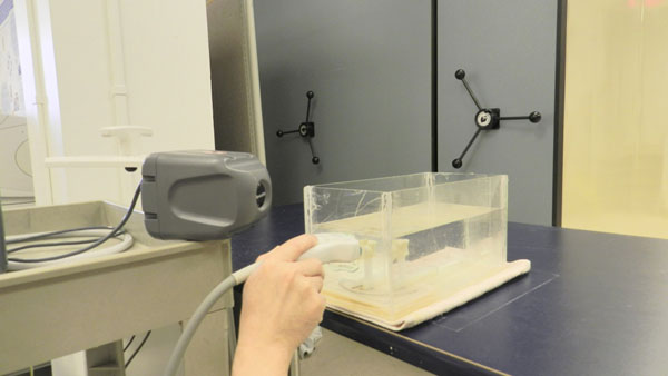
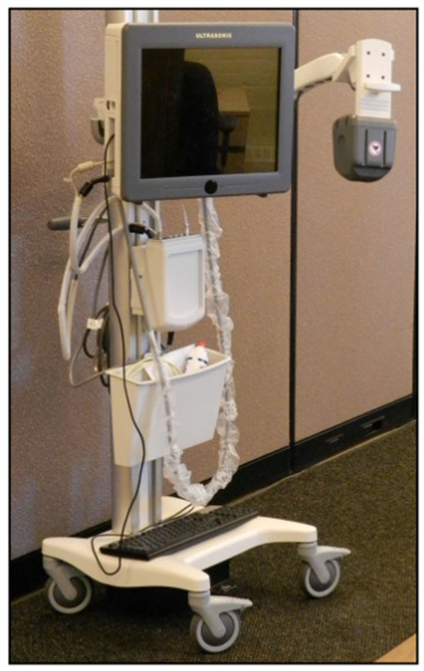
Methods: Three cadaveric vertebrae T7, L1, and L3 were scanned with a 3D medical ultrasound system. Nine sets of ultrasound data, the vertebral rotation from 0 to 40° with 5° increments, were recorded from each vertebra. An in-house program was developed to reconstruct and measure the 3D vertebral images. The rotation of each reconstructed vertebra was determined by the angle between the line going through the centres of either laminae (L-L) or transverse processes (TP-TP) and a reference vertical plane. Three raters measured the rotation in 3 sessions, in which they used the mouse pointer to select the L-L or TP-TP according to their knowledge of vertebral anatomy. The program detected the 3D coordinates of these points and calculated the AVR. The intra-class correlation coefficients (ICCs) were used to calculate the intra-reliability and inter-reliability. The mean absolute difference (MAD±SD) and the range of difference (RD) between the actual values and the average measurements of each rater were computed to evaluate the accuracy of methods.
Results: When rotation was greater than 30° for both L1 and L3, all raters found it difficult to determine one of the lamina areas due to the lack of ultrasound information in an area behind the spinous process. Therefore, the corresponding measurements were excluded. The ICC values of the intra-reliability (L-L, TP-TP) for the three raters were (0.987, 0.991), (0.989, 0.998) and (0.997, 1.000), respectively; meanwhile, the inter-reliability were 0.991 for (L-L) and 0.992 for (TP-TP). All ICC values were greater than 0.98 indicating both methods were highly reliable. The MAD±SD values (L-L, TP-TP) for the three raters were (1.5±0.3°, 1.2±0.2°), (1.6±0.3°, 1.3±0.3°), and (1.7±0.5°, 0.9±0.2°), respectively. The RD (L-L, TP-TP) were (0-4.5°, 0-3.5°), (0-5.1°, 0-4.3°), and (0-5.1°, 0-2.8°) for the three raters, respectively.
Conclusions: The (L-L) and (TP-TP) methods could be used to measure AVR reliability from the 3D ultrasound images.

 求助内容:
求助内容: 应助结果提醒方式:


