Matthias S Roost, Liesbeth van Iperen, Ana de Melo Bernardo, Christine L Mummery, Françoise Carlotti, Eelco Jp de Koning, Susana M Chuva de Sousa Lopes
{"title":"Lymphangiogenesis and angiogenesis during human fetal pancreas development.","authors":"Matthias S Roost, Liesbeth van Iperen, Ana de Melo Bernardo, Christine L Mummery, Françoise Carlotti, Eelco Jp de Koning, Susana M Chuva de Sousa Lopes","doi":"10.1186/2045-824X-6-22","DOIUrl":null,"url":null,"abstract":"<p><strong>Background: </strong>The complex endocrine and exocrine functionality of the human pancreas depends on an efficient fluid transport through the blood and the lymphatic vascular systems. The lymphatic vasculature has key roles in the physiology of the pancreas and in regulating the immune response, both important for developing successful transplantation and cell-replacement therapies to treat diabetes. However, little is known about how the lymphatic and blood systems develop in humans. Here, we investigated the establishment of these two vascular systems in human pancreas organogenesis in order to understand neovascularization in the context of emerging regenerative therapies.</p><p><strong>Methods: </strong>We examined angiogenesis and lymphangiogenesis during human pancreas development between 9 and 22 weeks of gestation (W9-W22) by immunohistochemistry.</p><p><strong>Results: </strong>As early as W9, the peri-pancreatic mesenchyme was populated by CD31-expressing blood vessels as well as LYVE1- and PDPN-expressing lymphatic vessels. The appearance of smooth muscle cell-coated blood vessels in the intra-pancreatic mesenchyme occurred only several weeks later and from W14.5 onwards the islets of Langerhans also became heavily irrigated by blood vessels. In contrast to blood vessels, LYVE1- and PDPN-expressing lymphatic vessels were restricted to the peri-pancreatic mesenchyme until later in development (W14.5-W17), and some of these invading lymphatic vessels contained smooth muscle cells at W17. Interestingly, between W11-W22, most large caliber lymphatic vessels were lined with a characteristic, discontinuous, collagen type IV-rich basement membrane. Whilst lymphatic vessels did not directly intrude the islets of Langerhans, three-dimensional reconstruction revealed that they were present in the vicinity of islets of Langerhans between W17-W22.</p><p><strong>Conclusion: </strong>Our data suggest that the blood and lymphatic machinery in the human pancreas is in place to support endocrine function from W17-W22 onwards. Our study provides the first systematic assessment of the progression of lymphangiogenesis during human pancreatic development.</p>","PeriodicalId":23948,"journal":{"name":"Vascular Cell","volume":"6 ","pages":"22"},"PeriodicalIF":0.0000,"publicationDate":"2014-11-01","publicationTypes":"Journal Article","fieldsOfStudy":null,"isOpenAccess":false,"openAccessPdf":"https://sci-hub-pdf.com/10.1186/2045-824X-6-22","citationCount":"18","resultStr":null,"platform":"Semanticscholar","paperid":null,"PeriodicalName":"Vascular Cell","FirstCategoryId":"1085","ListUrlMain":"https://doi.org/10.1186/2045-824X-6-22","RegionNum":0,"RegionCategory":null,"ArticlePicture":[],"TitleCN":null,"AbstractTextCN":null,"PMCID":null,"EPubDate":"2014/1/1 0:00:00","PubModel":"eCollection","JCR":"Q4","JCRName":"Neuroscience","Score":null,"Total":0}
引用次数: 18
Abstract
Background: The complex endocrine and exocrine functionality of the human pancreas depends on an efficient fluid transport through the blood and the lymphatic vascular systems. The lymphatic vasculature has key roles in the physiology of the pancreas and in regulating the immune response, both important for developing successful transplantation and cell-replacement therapies to treat diabetes. However, little is known about how the lymphatic and blood systems develop in humans. Here, we investigated the establishment of these two vascular systems in human pancreas organogenesis in order to understand neovascularization in the context of emerging regenerative therapies.
Methods: We examined angiogenesis and lymphangiogenesis during human pancreas development between 9 and 22 weeks of gestation (W9-W22) by immunohistochemistry.
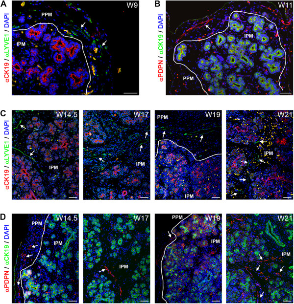
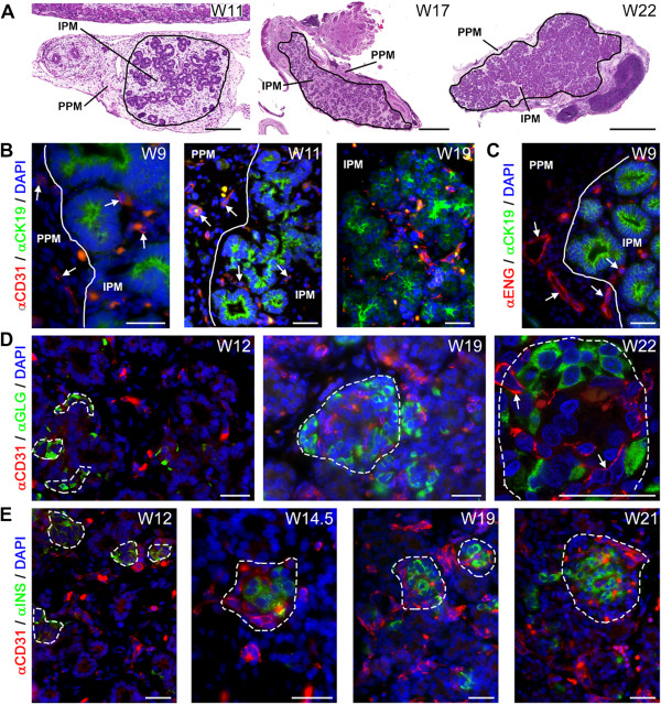
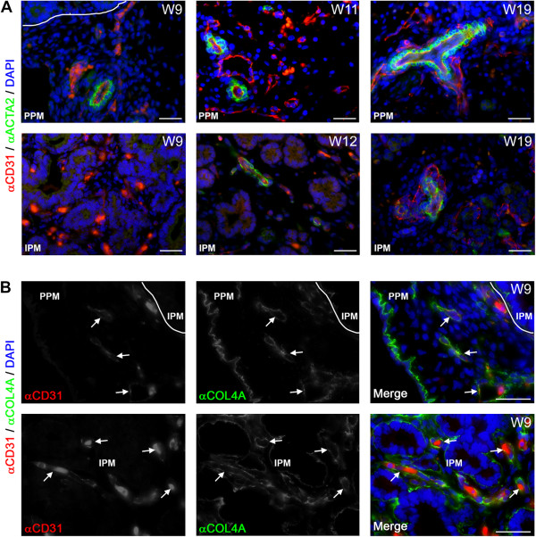
Results: As early as W9, the peri-pancreatic mesenchyme was populated by CD31-expressing blood vessels as well as LYVE1- and PDPN-expressing lymphatic vessels. The appearance of smooth muscle cell-coated blood vessels in the intra-pancreatic mesenchyme occurred only several weeks later and from W14.5 onwards the islets of Langerhans also became heavily irrigated by blood vessels. In contrast to blood vessels, LYVE1- and PDPN-expressing lymphatic vessels were restricted to the peri-pancreatic mesenchyme until later in development (W14.5-W17), and some of these invading lymphatic vessels contained smooth muscle cells at W17. Interestingly, between W11-W22, most large caliber lymphatic vessels were lined with a characteristic, discontinuous, collagen type IV-rich basement membrane. Whilst lymphatic vessels did not directly intrude the islets of Langerhans, three-dimensional reconstruction revealed that they were present in the vicinity of islets of Langerhans between W17-W22.
Conclusion: Our data suggest that the blood and lymphatic machinery in the human pancreas is in place to support endocrine function from W17-W22 onwards. Our study provides the first systematic assessment of the progression of lymphangiogenesis during human pancreatic development.

 求助内容:
求助内容: 应助结果提醒方式:


