John D Miller, Timothy M Rankin, Natalie T Hua, Tina Ontiveros, Nicholas A Giovinco, Joseph L Mills, David G Armstrong
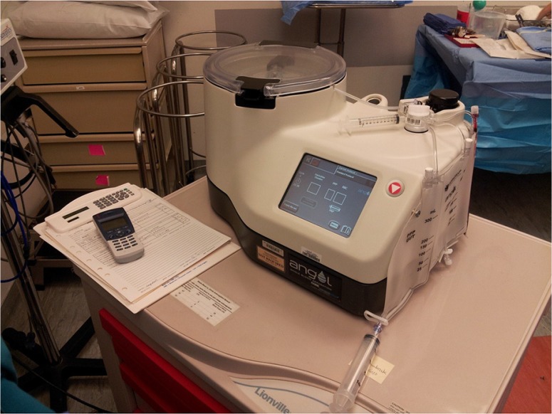
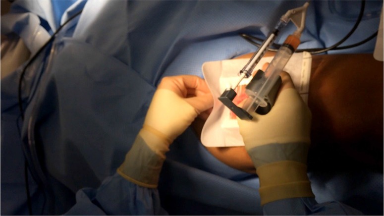
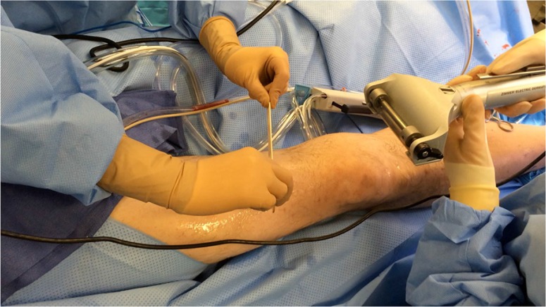
{"title":"Reduction of pain via platelet-rich plasma in split-thickness skin graft donor sites: a series of matched pairs.","authors":"John D Miller, Timothy M Rankin, Natalie T Hua, Tina Ontiveros, Nicholas A Giovinco, Joseph L Mills, David G Armstrong","doi":"10.3402/dfa.v6.24972","DOIUrl":null,"url":null,"abstract":"<p><p>In the past decade, autologous platelet-rich plasma (PRP) therapy has seen increasingly widespread integration into medical specialties. PRP application is known to accelerate wound epithelialization rates, and may also reduce postoperative wound site pain. Recently, we observed an increase in patient satisfaction following PRP gel (Angel, Cytomedix, Rockville, MD) application to split-thickness skin graft (STSG) donor sites. We assessed all patients known to our university-based hospital service who underwent multiple STSGs up to the year 2014, with at least one treated with topical PRP. Based on these criteria, five patients aged 48.4±17.6 (80% male) were identified who could serve as their own control, with mean time of 4.4±5.1 years between operations. In both therapies, initial dressing changes occurred on postoperative day (POD) 7, with donor site pain measured by Likert visual pain scale. Paired t-tests compared the size and thickness of harvested skin graft and patient pain level, and STSG thickness and surface area were comparable between control and PRP interventions (p>0.05 for all). Donor site pain was reduced from an average of 7.2 (±2.6) to 3 (±3.7), an average reduction in pain of 4.2 (standard error 1.1, p=0.0098) following PRP use. Based on these results, the authors suggest PRP as a beneficial adjunct for reducing donor site pain following STSG harvest. </p>","PeriodicalId":45385,"journal":{"name":"Diabetic Foot & Ankle","volume":"6 ","pages":"24972"},"PeriodicalIF":0.0000,"publicationDate":"2015-01-22","publicationTypes":"Journal Article","fieldsOfStudy":null,"isOpenAccess":false,"openAccessPdf":"https://sci-hub-pdf.com/10.3402/dfa.v6.24972","citationCount":"23","resultStr":null,"platform":"Semanticscholar","paperid":null,"PeriodicalName":"Diabetic Foot & Ankle","FirstCategoryId":"1085","ListUrlMain":"https://doi.org/10.3402/dfa.v6.24972","RegionNum":0,"RegionCategory":null,"ArticlePicture":[],"TitleCN":null,"AbstractTextCN":null,"PMCID":null,"EPubDate":"2015/1/1 0:00:00","PubModel":"eCollection","JCR":"Q1","JCRName":"Health Professions","Score":null,"Total":0}
引用次数: 23
Abstract
In the past decade, autologous platelet-rich plasma (PRP) therapy has seen increasingly widespread integration into medical specialties. PRP application is known to accelerate wound epithelialization rates, and may also reduce postoperative wound site pain. Recently, we observed an increase in patient satisfaction following PRP gel (Angel, Cytomedix, Rockville, MD) application to split-thickness skin graft (STSG) donor sites. We assessed all patients known to our university-based hospital service who underwent multiple STSGs up to the year 2014, with at least one treated with topical PRP. Based on these criteria, five patients aged 48.4±17.6 (80% male) were identified who could serve as their own control, with mean time of 4.4±5.1 years between operations. In both therapies, initial dressing changes occurred on postoperative day (POD) 7, with donor site pain measured by Likert visual pain scale. Paired t-tests compared the size and thickness of harvested skin graft and patient pain level, and STSG thickness and surface area were comparable between control and PRP interventions (p>0.05 for all). Donor site pain was reduced from an average of 7.2 (±2.6) to 3 (±3.7), an average reduction in pain of 4.2 (standard error 1.1, p=0.0098) following PRP use. Based on these results, the authors suggest PRP as a beneficial adjunct for reducing donor site pain following STSG harvest.

 求助内容:
求助内容: 应助结果提醒方式:


