Neph1 is reduced in primary focal segmental glomerulosclerosis, minimal change nephrotic syndrome, and corresponding experimental animal models of adriamycin-induced nephropathy and puromycin aminonucleoside nephrosis.
Jenny Hulkko, Jaakko Patrakka, Mark Lal, Karl Tryggvason, Kjell Hultenby, Annika Wernerson
{"title":"Neph1 is reduced in primary focal segmental glomerulosclerosis, minimal change nephrotic syndrome, and corresponding experimental animal models of adriamycin-induced nephropathy and puromycin aminonucleoside nephrosis.","authors":"Jenny Hulkko, Jaakko Patrakka, Mark Lal, Karl Tryggvason, Kjell Hultenby, Annika Wernerson","doi":"10.1159/000365091","DOIUrl":null,"url":null,"abstract":"<p><strong>Background/aims: </strong>The transmembrane proteins Neph1 and nephrin form a complex in the slit diaphragm (SD) of podocytes. As recent studies indicate an involvement of this complex in the polymerization of the actin cytoskeleton and proteinuria, we wanted to study the subcellular localization of Neph1 in the normal human kidney and its expression in focal segmental glomerulosclerosis (FSGS), minimal change nephrotic syndrome (MCNS), and the corresponding experimental models of Adriamycin-induced nephropathy (ADR) and puromycin aminonucleoside nephrosis (PAN). All these disorders are characterized by substantial foot process effacement (FPE) and proteinuria.</p><p><strong>Materials and methods: </strong>Kidney biopsies from patients with primary FSGS (perihilar type) and MCNS were compared to normal renal tissue. Mouse and rat kidney cortices from days 7 and 14 after Adriamycin injection and days 2 and 4 after puromycin aminonucleoside injection, respectively, were compared to control mouse and rat kidney. Polyclonal antibodies against Neph1 and nephrin were used for immunoelectron microscopy, and semiquantification was performed.</p><p><strong>Results: </strong>We localized Neph1 mainly to, and in close proximity to, the SD. Double staining of Neph1 and nephrin showed the proteins to be in close connection in the SD. The total amount of Neph1 in the podocytes was significantly reduced in FSGS, MCNS, ADR, and PAN. The reduction of Neph1 was also seen in areas with and without FPE. Nephrin was reduced in MCNS and PAN but unchanged in FSGS.</p><p><strong>Conclusion: </strong>With nephrin (but not Neph1) unchanged in FSGS, there might be a disruption of the complex and an involvement of Neph1 in its pathogenesis.</p>","PeriodicalId":56356,"journal":{"name":"Nephron Extra","volume":"4 3","pages":"146-54"},"PeriodicalIF":0.0000,"publicationDate":"2014-09-19","publicationTypes":"Journal Article","fieldsOfStudy":null,"isOpenAccess":false,"openAccessPdf":"https://sci-hub-pdf.com/10.1159/000365091","citationCount":"13","resultStr":null,"platform":"Semanticscholar","paperid":null,"PeriodicalName":"Nephron Extra","FirstCategoryId":"1085","ListUrlMain":"https://doi.org/10.1159/000365091","RegionNum":0,"RegionCategory":null,"ArticlePicture":[],"TitleCN":null,"AbstractTextCN":null,"PMCID":null,"EPubDate":"2014/9/1 0:00:00","PubModel":"eCollection","JCR":"","JCRName":"","Score":null,"Total":0}
引用次数: 13
Abstract
Background/aims: The transmembrane proteins Neph1 and nephrin form a complex in the slit diaphragm (SD) of podocytes. As recent studies indicate an involvement of this complex in the polymerization of the actin cytoskeleton and proteinuria, we wanted to study the subcellular localization of Neph1 in the normal human kidney and its expression in focal segmental glomerulosclerosis (FSGS), minimal change nephrotic syndrome (MCNS), and the corresponding experimental models of Adriamycin-induced nephropathy (ADR) and puromycin aminonucleoside nephrosis (PAN). All these disorders are characterized by substantial foot process effacement (FPE) and proteinuria.
Materials and methods: Kidney biopsies from patients with primary FSGS (perihilar type) and MCNS were compared to normal renal tissue. Mouse and rat kidney cortices from days 7 and 14 after Adriamycin injection and days 2 and 4 after puromycin aminonucleoside injection, respectively, were compared to control mouse and rat kidney. Polyclonal antibodies against Neph1 and nephrin were used for immunoelectron microscopy, and semiquantification was performed.
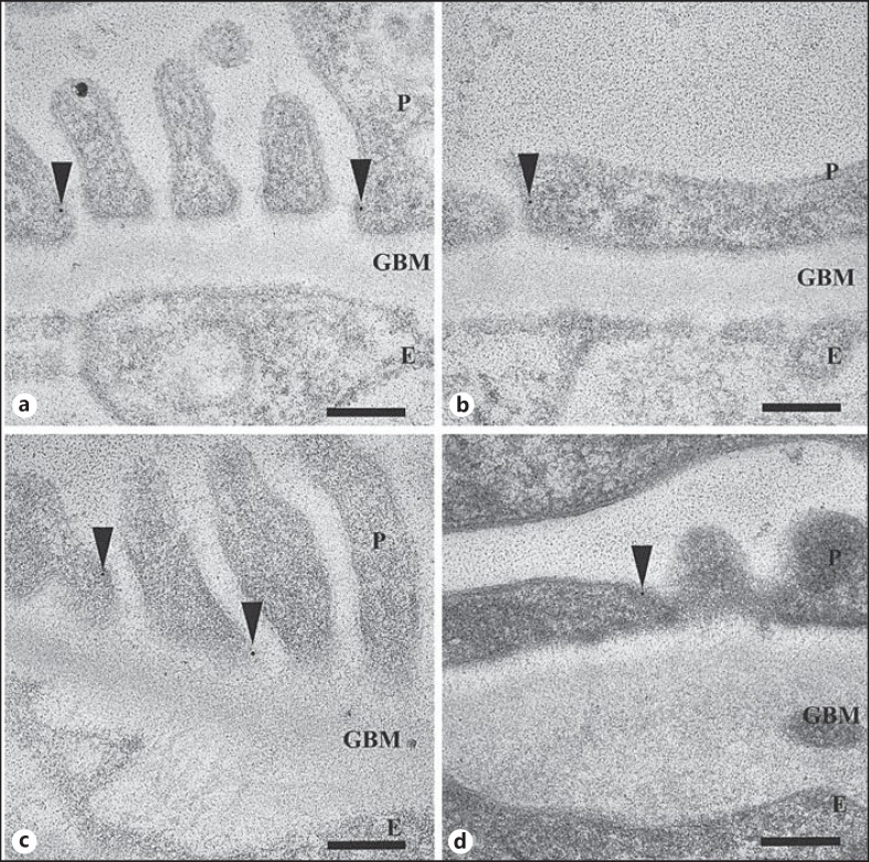
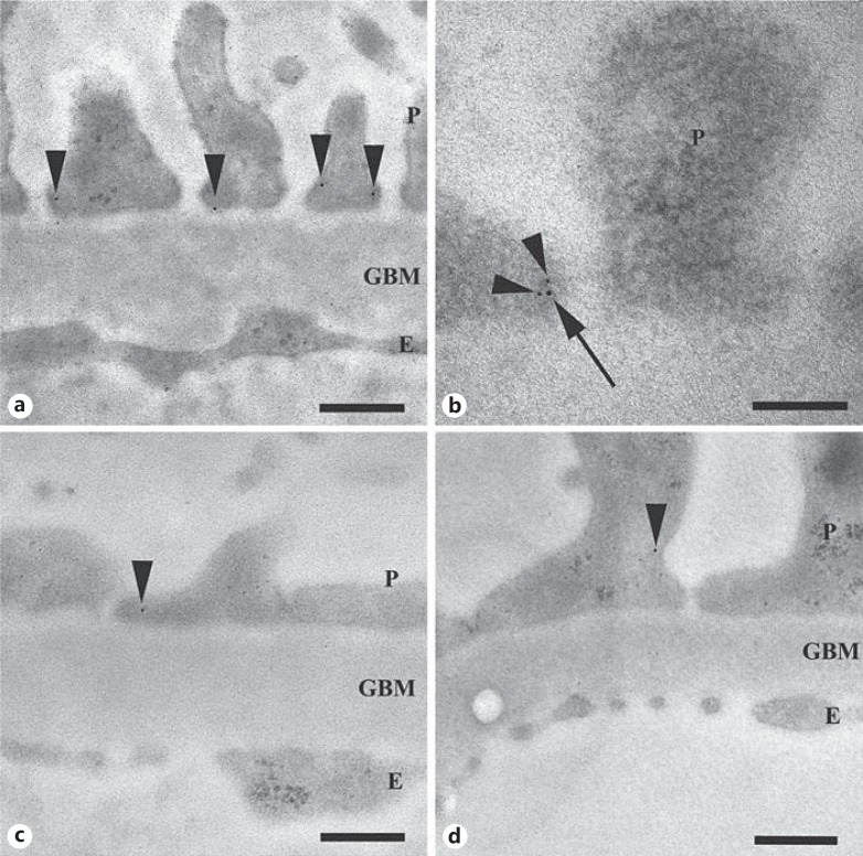
Results: We localized Neph1 mainly to, and in close proximity to, the SD. Double staining of Neph1 and nephrin showed the proteins to be in close connection in the SD. The total amount of Neph1 in the podocytes was significantly reduced in FSGS, MCNS, ADR, and PAN. The reduction of Neph1 was also seen in areas with and without FPE. Nephrin was reduced in MCNS and PAN but unchanged in FSGS.
Conclusion: With nephrin (but not Neph1) unchanged in FSGS, there might be a disruption of the complex and an involvement of Neph1 in its pathogenesis.
期刊介绍:
An open-access subjournal to Nephron. ''Nephron EXTRA'' publishes additional high-quality articles that cannot be published in the main journal ''Nephron'' due to space limitations.

 求助内容:
求助内容: 应助结果提醒方式:


