{"title":"Extracellular ATP does not induce P2X7 receptor-dependent responses in cultured renal- and liver-derived swine macrophages","authors":"Takato Takenouchi , Shunichi Suzuki , Hiroki Shinkai , Mitsutoshi Tsukimoto , Mitsuru Sato , Hirohide Uenishi , Hiroshi Kitani","doi":"10.1016/j.rinim.2014.07.002","DOIUrl":null,"url":null,"abstract":"<div><p>The P2X7 receptor (P2X7R) is an ATP-gated cation channel that is abundantly expressed in monocytes/macrophages. P2X7R activation by ATP results in various cellular responses including Ca<sup>2+</sup> influx, membrane pore formation, and cytokine secretion. Since P2X7R has low affinity for ATP, high concentrations of ATP (in the mM range) are generally required to activate this receptor <em>in vitro</em>. Functional expression of P2X7R has been detected in monocytes/macrophages obtained from different animal species including humans, rodents, dogs, and bovines, but so far it has not been detected in swine (<em>Sus scrofa</em>). In this study, we investigated the expression and functions of P2X7R in swine macrophages, which were isolated from mixed primary cultures of swine kidney or liver tissue. The P2X7R mRNA and protein expression observed in the swine macrophages was comparable to that seen in a <em>c-myc</em>-immortalized mouse kidney-derived clonal macrophage cell line (KM-1). However, extracellular ATP did not induce P2X7R-dependent sustained Ca<sup>2+</sup> influx, membrane pore formation, or the secretion of the bioactive cytokine interleukin-1β in the swine macrophages, whereas these responses were clearly observed in the mouse KM-1 cells after stimulation with millimolar concentrations of ATP as a positive control. These findings suggest that the ATP/P2X7R pathway is impaired in swine macrophages at least in the culture conditions used in the present study.</p></div>","PeriodicalId":89845,"journal":{"name":"Results in immunology","volume":"4 ","pages":"Pages 62-67"},"PeriodicalIF":0.0000,"publicationDate":"2014-01-01","publicationTypes":"Journal Article","fieldsOfStudy":null,"isOpenAccess":false,"openAccessPdf":"https://sci-hub-pdf.com/10.1016/j.rinim.2014.07.002","citationCount":"17","resultStr":null,"platform":"Semanticscholar","paperid":null,"PeriodicalName":"Results in immunology","FirstCategoryId":"1085","ListUrlMain":"https://www.sciencedirect.com/science/article/pii/S2211283914000094","RegionNum":0,"RegionCategory":null,"ArticlePicture":[],"TitleCN":null,"AbstractTextCN":null,"PMCID":null,"EPubDate":"","PubModel":"","JCR":"","JCRName":"","Score":null,"Total":0}
引用次数: 17
Abstract
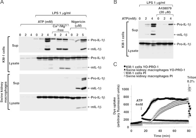
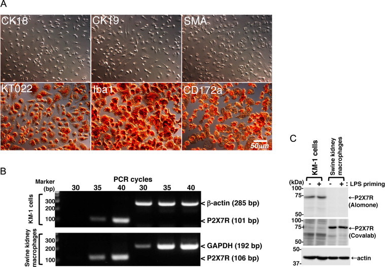
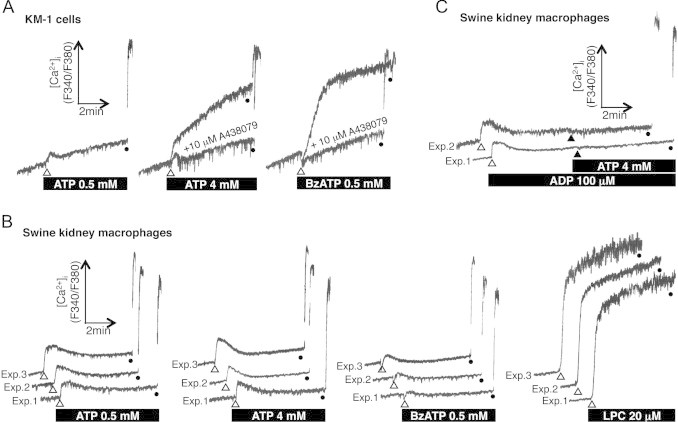
The P2X7 receptor (P2X7R) is an ATP-gated cation channel that is abundantly expressed in monocytes/macrophages. P2X7R activation by ATP results in various cellular responses including Ca2+ influx, membrane pore formation, and cytokine secretion. Since P2X7R has low affinity for ATP, high concentrations of ATP (in the mM range) are generally required to activate this receptor in vitro. Functional expression of P2X7R has been detected in monocytes/macrophages obtained from different animal species including humans, rodents, dogs, and bovines, but so far it has not been detected in swine (Sus scrofa). In this study, we investigated the expression and functions of P2X7R in swine macrophages, which were isolated from mixed primary cultures of swine kidney or liver tissue. The P2X7R mRNA and protein expression observed in the swine macrophages was comparable to that seen in a c-myc-immortalized mouse kidney-derived clonal macrophage cell line (KM-1). However, extracellular ATP did not induce P2X7R-dependent sustained Ca2+ influx, membrane pore formation, or the secretion of the bioactive cytokine interleukin-1β in the swine macrophages, whereas these responses were clearly observed in the mouse KM-1 cells after stimulation with millimolar concentrations of ATP as a positive control. These findings suggest that the ATP/P2X7R pathway is impaired in swine macrophages at least in the culture conditions used in the present study.

 求助内容:
求助内容: 应助结果提醒方式:


