Cristan Herbert, Qing-Xiang Zeng, Ramesh Shanmugasundaram, Linda Garthwaite, Brian G Oliver, Rakesh K Kumar
{"title":"Response of airway epithelial cells to double-stranded RNA in an allergic environment.","authors":"Cristan Herbert, Qing-Xiang Zeng, Ramesh Shanmugasundaram, Linda Garthwaite, Brian G Oliver, Rakesh K Kumar","doi":"10.1186/s40247-014-0011-6","DOIUrl":null,"url":null,"abstract":"<p><strong>Background: </strong>Respiratory viral infections are the most common trigger of acute exacerbations in patients with allergic asthma. The anti-viral response of airway epithelial cells (AEC) may be impaired in asthmatics, while cytokines produced by AEC may drive the inflammatory response. We investigated whether AEC cultured in the presence of Th2 cytokines associated with an allergic environment exhibited altered responses to double-stranded RNA, a virus-like stimulus.</p><p><strong>Methods: </strong>We undertook preliminary studies using the MLE-12 cell line derived from mouse distal respiratory epithelial cells, then confirmed and extended our findings using low-passage human AEC. Cells were cultured in the absence or presence of the Th2 cytokines IL-4 and IL-13 for 48 hours, then stimulated with poly I:C for 4 hours. Expression of relevant anti-viral response and cytokine genes was assessed by quantitative real-time PCR. Secretion of cytokine proteins was assessed by immunoassay.</p><p><strong>Results: </strong>Following stimulation with poly I:C, MLE-12 cells pre-treated with Th2 cytokines exhibited significantly higher levels of expression of mRNA for the cytokine genes Cxcl10 and Cxcl11, as well as a trend towards increased expression of Cxcl9 and Il6. Expression of anti-viral response genes was mostly unchanged, although Stat1, Ifit1 and Ifitm3 were significantly increased in Th2 cytokine pre-treated cells. Human AEC pre-treated with IL-4 and IL-13, then stimulated with poly I:C, similarly exhibited significantly higher expression of IL8, CXCL9, CXCL10, CXCL11 and CCL5 genes. In parallel, there was significantly increased secretion of CXCL8 and CCL5, as well as a trend towards increased secretion of CXCL10 and IL-6. Again, expression of anti-viral response genes was not decreased. Rather, there was significantly enhanced expression of mRNA for type III interferons, RNA helicases and other interferon-stimulated genes.</p><p><strong>Conclusion: </strong>The Th2 cytokine environment appears to promote increased production of pro-inflammatory chemokines by AEC in response to double-stranded RNA, which could help explain the exaggerated inflammatory response to respiratory viral infection in allergic asthmatics. However, any impairment of anti-viral host defences in asthmatics appears unlikely to be a consequence of Th2 cytokine-induced downregulation of the expression of viral response genes by AEC.</p>","PeriodicalId":90074,"journal":{"name":"Translational respiratory medicine","volume":"2 1","pages":"11"},"PeriodicalIF":0.0000,"publicationDate":"2014-12-01","publicationTypes":"Journal Article","fieldsOfStudy":null,"isOpenAccess":false,"openAccessPdf":"https://sci-hub-pdf.com/10.1186/s40247-014-0011-6","citationCount":"17","resultStr":null,"platform":"Semanticscholar","paperid":null,"PeriodicalName":"Translational respiratory medicine","FirstCategoryId":"1085","ListUrlMain":"https://doi.org/10.1186/s40247-014-0011-6","RegionNum":0,"RegionCategory":null,"ArticlePicture":[],"TitleCN":null,"AbstractTextCN":null,"PMCID":null,"EPubDate":"2014/9/11 0:00:00","PubModel":"Epub","JCR":"","JCRName":"","Score":null,"Total":0}
引用次数: 17
Abstract
Background: Respiratory viral infections are the most common trigger of acute exacerbations in patients with allergic asthma. The anti-viral response of airway epithelial cells (AEC) may be impaired in asthmatics, while cytokines produced by AEC may drive the inflammatory response. We investigated whether AEC cultured in the presence of Th2 cytokines associated with an allergic environment exhibited altered responses to double-stranded RNA, a virus-like stimulus.
Methods: We undertook preliminary studies using the MLE-12 cell line derived from mouse distal respiratory epithelial cells, then confirmed and extended our findings using low-passage human AEC. Cells were cultured in the absence or presence of the Th2 cytokines IL-4 and IL-13 for 48 hours, then stimulated with poly I:C for 4 hours. Expression of relevant anti-viral response and cytokine genes was assessed by quantitative real-time PCR. Secretion of cytokine proteins was assessed by immunoassay.
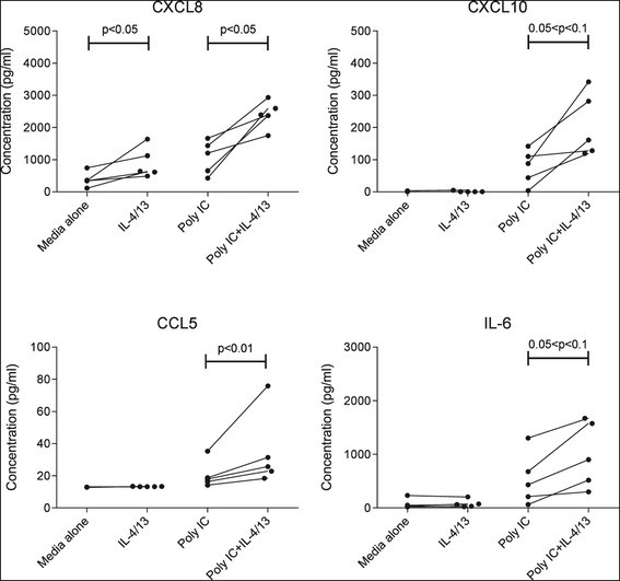
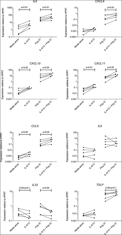
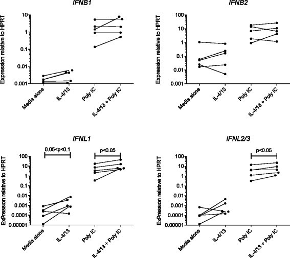
Results: Following stimulation with poly I:C, MLE-12 cells pre-treated with Th2 cytokines exhibited significantly higher levels of expression of mRNA for the cytokine genes Cxcl10 and Cxcl11, as well as a trend towards increased expression of Cxcl9 and Il6. Expression of anti-viral response genes was mostly unchanged, although Stat1, Ifit1 and Ifitm3 were significantly increased in Th2 cytokine pre-treated cells. Human AEC pre-treated with IL-4 and IL-13, then stimulated with poly I:C, similarly exhibited significantly higher expression of IL8, CXCL9, CXCL10, CXCL11 and CCL5 genes. In parallel, there was significantly increased secretion of CXCL8 and CCL5, as well as a trend towards increased secretion of CXCL10 and IL-6. Again, expression of anti-viral response genes was not decreased. Rather, there was significantly enhanced expression of mRNA for type III interferons, RNA helicases and other interferon-stimulated genes.
Conclusion: The Th2 cytokine environment appears to promote increased production of pro-inflammatory chemokines by AEC in response to double-stranded RNA, which could help explain the exaggerated inflammatory response to respiratory viral infection in allergic asthmatics. However, any impairment of anti-viral host defences in asthmatics appears unlikely to be a consequence of Th2 cytokine-induced downregulation of the expression of viral response genes by AEC.

 求助内容:
求助内容: 应助结果提醒方式:


