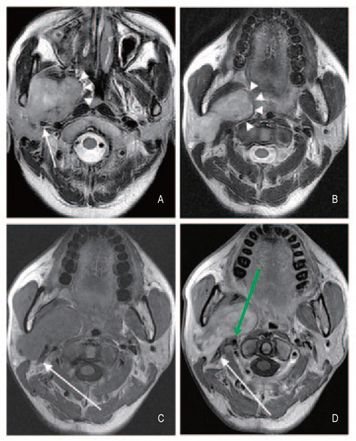
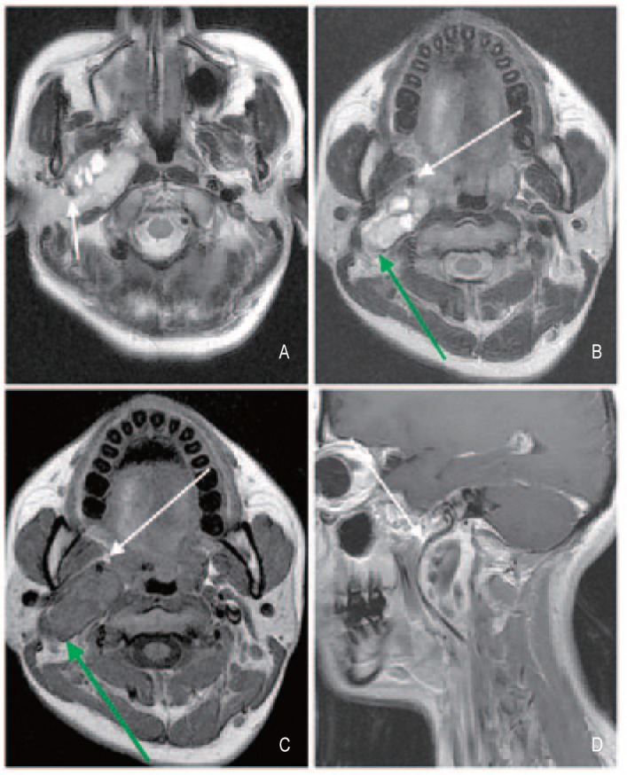
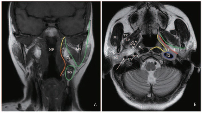
{"title":"A modified method for locating parapharyngeal space neoplasms on magnetic resonance images: implications for differential diagnosis.","authors":"Xue-Wen Liu, Ling Wang, Hui Li, Rong Zhang, Zhi-Jun Geng, De-Ling Wang, Chuan-Miao Xie","doi":"10.5732/cjc.014.10017","DOIUrl":null,"url":null,"abstract":"<p><p>The parapharyngeal space (PPS) is an inverted pyramid-shaped deep space in the head and neck region, and a variety of tumors, such as salivary gland tumors, neurogenic tumors, nasopharyngeal carcinomas with parapharyngeal invasion, and lymphomas, can be found in this space. The differential diagnosis of PPS tumors remains challenging for radiologists. This study aimed to develop and test a modified method for locating PPS tumors on magnetic resonance (MR) images to improve preoperative differential diagnosis. The new protocol divided the PPS into three compartments: a prestyloid compartment, the carotid sheath, and the areas outside the carotid sheath. PPS tumors were located in these compartments according to the displacements of the tensor veli palatini muscle and the styloid process, with or without blood vessel separations and medial pterygoid invasion. This protocol, as well as a more conventional protocol that is based on displacements of the internal carotid artery (ICA), was used to assess MR images captured from a series of 58 PPS tumors. The consequent distributions of PPS tumor locations determined by both methods were compared. Of all 58 tumors, our new method determined that 57 could be assigned to precise PPS compartments. Nearly all (13/14; 93%) tumors that were located in the pre-styloid compartment were salivary gland tumors. All 15 tumors within the carotid sheath were neurogenic tumors. The vast majority (18/20; 90%) of trans-spatial lesions were malignancies. However, according to the ICA-based method, 28 tumors were located in the pre-styloid compartment, and 24 were located in the post-styloid compartment, leaving 6 tumors that were difficult to locate. Lesions located in both the pre-styloid and the post-styloid compartments comprised various types of tumors. Compared with the conventional ICA-based method, our new method can help radiologists to narrow the differential diagnosis of PPS tumors to specific compartments. </p>","PeriodicalId":10034,"journal":{"name":"癌症","volume":"33 10","pages":"511-20"},"PeriodicalIF":0.0000,"publicationDate":"2014-10-01","publicationTypes":"Journal Article","fieldsOfStudy":null,"isOpenAccess":false,"openAccessPdf":"https://ftp.ncbi.nlm.nih.gov/pub/pmc/oa_pdf/0b/5d/cjc-33-10-511.PMC4198754.pdf","citationCount":"11","resultStr":null,"platform":"Semanticscholar","paperid":null,"PeriodicalName":"癌症","FirstCategoryId":"3","ListUrlMain":"https://doi.org/10.5732/cjc.014.10017","RegionNum":0,"RegionCategory":null,"ArticlePicture":[],"TitleCN":null,"AbstractTextCN":null,"PMCID":null,"EPubDate":"2014/8/5 0:00:00","PubModel":"Epub","JCR":"Q","JCRName":"Medicine","Score":null,"Total":0}
引用次数: 11
Abstract
The parapharyngeal space (PPS) is an inverted pyramid-shaped deep space in the head and neck region, and a variety of tumors, such as salivary gland tumors, neurogenic tumors, nasopharyngeal carcinomas with parapharyngeal invasion, and lymphomas, can be found in this space. The differential diagnosis of PPS tumors remains challenging for radiologists. This study aimed to develop and test a modified method for locating PPS tumors on magnetic resonance (MR) images to improve preoperative differential diagnosis. The new protocol divided the PPS into three compartments: a prestyloid compartment, the carotid sheath, and the areas outside the carotid sheath. PPS tumors were located in these compartments according to the displacements of the tensor veli palatini muscle and the styloid process, with or without blood vessel separations and medial pterygoid invasion. This protocol, as well as a more conventional protocol that is based on displacements of the internal carotid artery (ICA), was used to assess MR images captured from a series of 58 PPS tumors. The consequent distributions of PPS tumor locations determined by both methods were compared. Of all 58 tumors, our new method determined that 57 could be assigned to precise PPS compartments. Nearly all (13/14; 93%) tumors that were located in the pre-styloid compartment were salivary gland tumors. All 15 tumors within the carotid sheath were neurogenic tumors. The vast majority (18/20; 90%) of trans-spatial lesions were malignancies. However, according to the ICA-based method, 28 tumors were located in the pre-styloid compartment, and 24 were located in the post-styloid compartment, leaving 6 tumors that were difficult to locate. Lesions located in both the pre-styloid and the post-styloid compartments comprised various types of tumors. Compared with the conventional ICA-based method, our new method can help radiologists to narrow the differential diagnosis of PPS tumors to specific compartments.
期刊介绍:
In July 2008, Landes Bioscience and Sun Yat-sen University Cancer Center began co-publishing the international, English-language version of AI ZHENG or the Chinese Journal of Cancer (CJC). CJC publishes original research, reviews, extra views, perspectives, supplements, and spotlights in all areas of cancer research. The primary criteria for publication in CJC are originality, outstanding scientific merit, and general interest. The Editorial Board is composed of members from around the world, who will strive to maintain the highest standards for excellence in order to generate a valuable resource for an international readership.
 求助内容:
求助内容: 应助结果提醒方式:


