Ultrasonographic classification of achilles tendon ruptures as a rationale for individual treatment selection.
引用次数: 36
Abstract
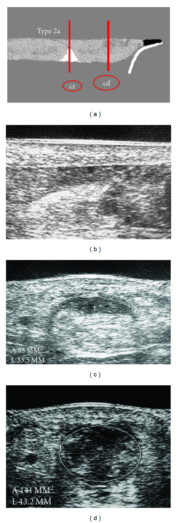
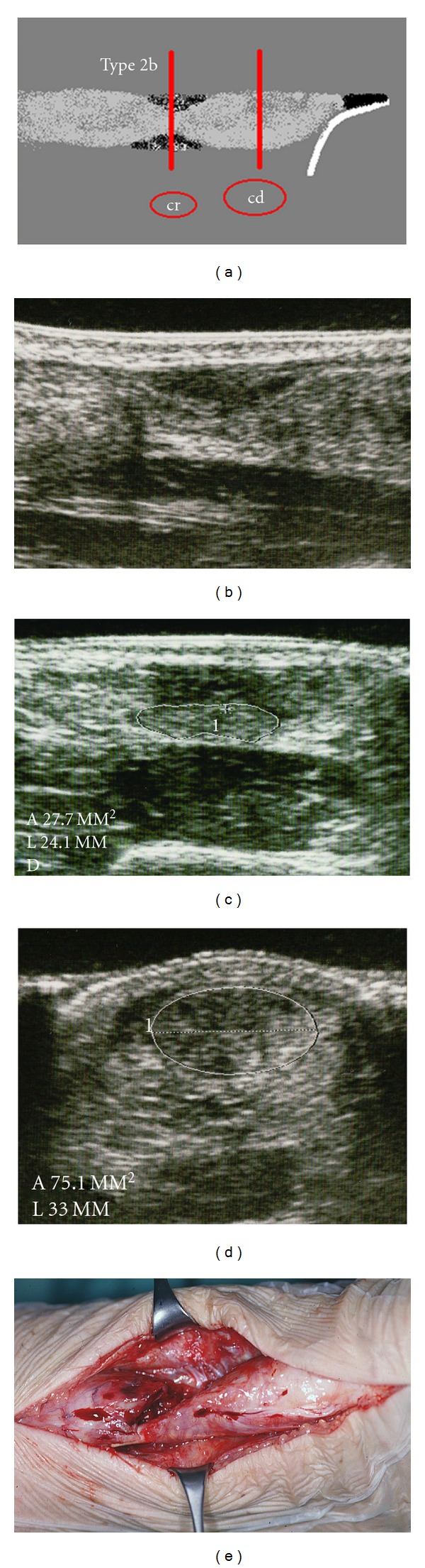
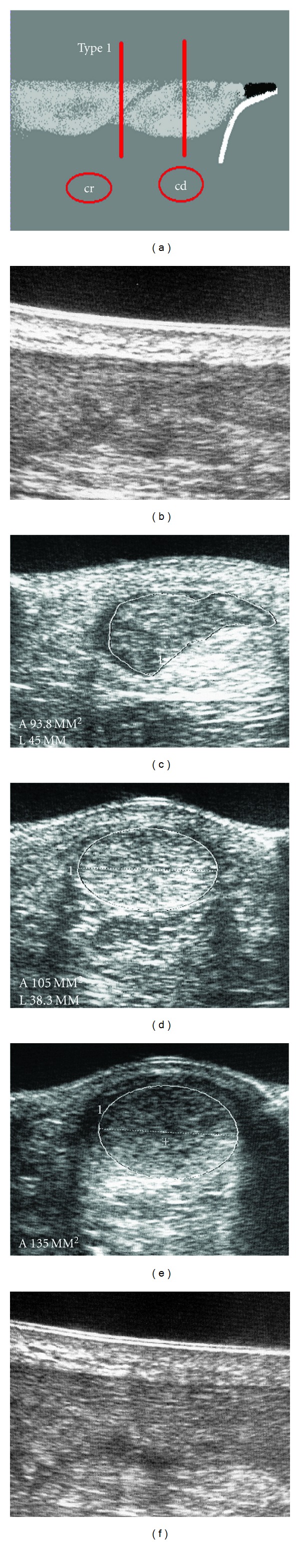
Purpose. This work introduces a distinct sonographic classification of Achilles tendon ruptures which has proven itself to be a reliable instrument for an individualized and differentiated therapy selection for patients who have suffered an Achilles tendon rupture. Materials and Methods. From January 1, 2000 to December 31, 2005, 273 patients who suffered from a complete subcutaneous rupture of the Achilles tendon (ASR) were clinically and sonographically evaluated. The sonographic classification was organized according to the location of the rupture, the contact of the tendon ends, and the structure of the interposition between the tendon ends. Results. In 266 of 273 (97.4%) patients the sonographic classification of the rupture of the Achilles tendon was recorded. Type 1 was detected in 54 patients (19.8%), type 2a in 68 (24.9%), type 2b in 33 (12.1%), type 3a in 20 (7.3%), type 3b in 61 (22.3%), type 4 in 20 (7.3%), and type 5 in 10 (3.7%). Of the patients with type 1 and fresh ASR, 96% (n = 47) were treated nonoperative-functionally, and 4% (n = 2) were treated by percutaneous suture with the Dresden instrument (pDI suture). Of the patients classified as type 2a with fresh ASR, 31 patients (48%) were treated nonoperatively-functionally and 33 patients (52%) with percutaneous suture with the Dresden instrument (pDI suture). Of the patients with type 3b and fresh ASR, 94% (n = 34) were treated by pDI suture and 6% (n = 2) by open suture according to Kirchmayr and Kessler. Conclusion. Unlike the clinical classification of the Achilles tendon rupture, the sonographic classification is a guide for deriving a graded and differentiated therapy from a broad spectrum of treatments.
跟腱断裂的超声分类作为个体治疗选择的依据。
目的。这项工作介绍了一种独特的跟腱断裂超声分类,它已被证明是一种可靠的工具,可以为患有跟腱断裂的患者提供个性化和差异化的治疗选择。材料与方法。从2000年1月1日至2005年12月31日,我们对273例完全性跟腱皮下断裂(ASR)患者进行了临床和超声检查。根据断裂部位、腱端接触情况及腱端间置结构进行超声分类。结果。273例患者中有266例(97.4%)记录了跟腱断裂的超声分型。1型54例(19.8%),2a型68例(24.9%),2b型33例(12.1%),3a型20例(7.3%),3b型61例(22.3%),4型20例(7.3%),5型10例(3.7%)。在1型和新鲜ASR患者中,96% (n = 47)采用非手术功能治疗,4% (n = 2)采用德累斯顿器械(pDI)经皮缝合治疗。在新鲜ASR的2a型患者中,31例(48%)患者采用非手术功能治疗,33例(52%)患者采用德累斯顿器械(pDI)经皮缝合。根据Kirchmayr和Kessler,在3b型和新鲜ASR患者中,94% (n = 34)采用pDI缝合,6% (n = 2)采用开放缝合。结论。与跟腱断裂的临床分类不同,超声分类是从广泛的治疗方法中得出分级和分化治疗的指南。
本文章由计算机程序翻译,如有差异,请以英文原文为准。
求助全文
约1分钟内获得全文
求助全文

 求助内容:
求助内容: 应助结果提醒方式:


