Ruken Yuksekkaya, Levent Aggunlu, Yusuf Oner, Halil Celik, Sergin Akpek, Fatih Celikyay
{"title":"Assessment of t2-weighted coronal magnetic resonance images in the investigation of pituitary lesions.","authors":"Ruken Yuksekkaya, Levent Aggunlu, Yusuf Oner, Halil Celik, Sergin Akpek, Fatih Celikyay","doi":"10.1155/2014/650926","DOIUrl":null,"url":null,"abstract":"<p><p>Magnetic resonance imaging is the most important diagnostic method in the investigation of the pituitary lesions. Our aim is to determine whether T2-weighted coronal images may be helpful in the evaluation of the pituitary gland with suspected pituitary adenomas. One hundred and sixty-seven patients were examined prospectively with T2-weighted coronal and T1-weighted coronal images enhanced with intravenous contrast material. The images were evaluated for the presence, the size, the location, and the ancillary signs including sellar floor erosion or ballooning, infindibulary deviation, convexity of the superior border of the gland, diffuse enlargement of the gland, and the invasion of the cavenous sinuses on both images. In forty-six (28%) patients lesions were revealed on both sequences. In twenty-one (12%) patients the lesions that were revealed on the T1-weighted images were not detected on the T2-weighted images. Positive predictive value, negative predictive value, sensitivity, specificity, and diagnostic accuracy rates of T2-weighted coronal images on the detection of the presence of lesions were 100%, 17.4%, 68.7%, 100%, and 87.4%, respectively. Both T2-weighted coronal and T1-weighted coronal images enhanced with intravenous gadolinium-based contrast material are important in the diagnosis of pituitary adenomas. T2-weighted coronal images could be used as a screening tool for the primary evaluation of the pituitary gland. </p>","PeriodicalId":90195,"journal":{"name":"ISRN radiology","volume":"2014 ","pages":"650926"},"PeriodicalIF":0.0000,"publicationDate":"2014-03-23","publicationTypes":"Journal Article","fieldsOfStudy":null,"isOpenAccess":false,"openAccessPdf":"https://sci-hub-pdf.com/10.1155/2014/650926","citationCount":"1","resultStr":null,"platform":"Semanticscholar","paperid":null,"PeriodicalName":"ISRN radiology","FirstCategoryId":"1085","ListUrlMain":"https://doi.org/10.1155/2014/650926","RegionNum":0,"RegionCategory":null,"ArticlePicture":[],"TitleCN":null,"AbstractTextCN":null,"PMCID":null,"EPubDate":"2014/1/1 0:00:00","PubModel":"eCollection","JCR":"","JCRName":"","Score":null,"Total":0}
引用次数: 1
Abstract
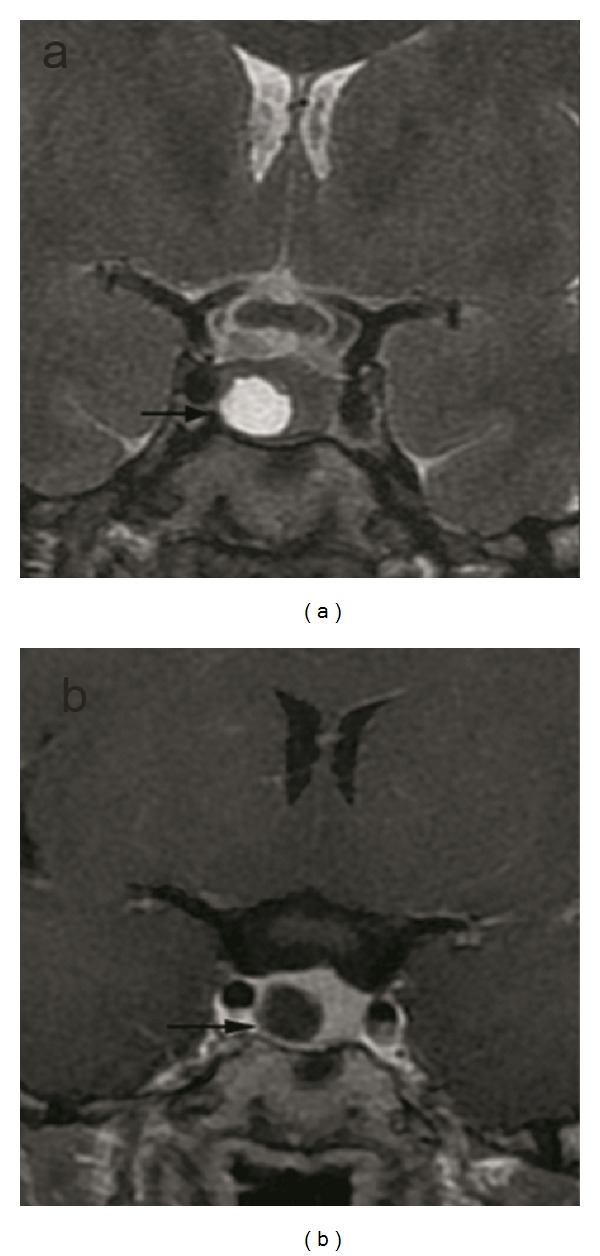
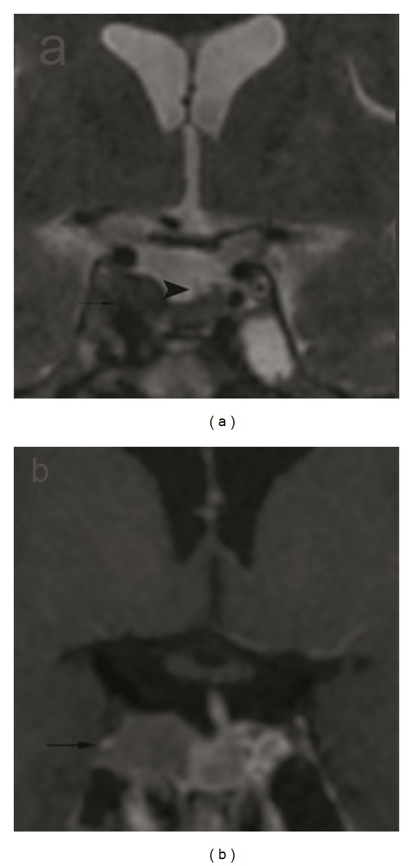
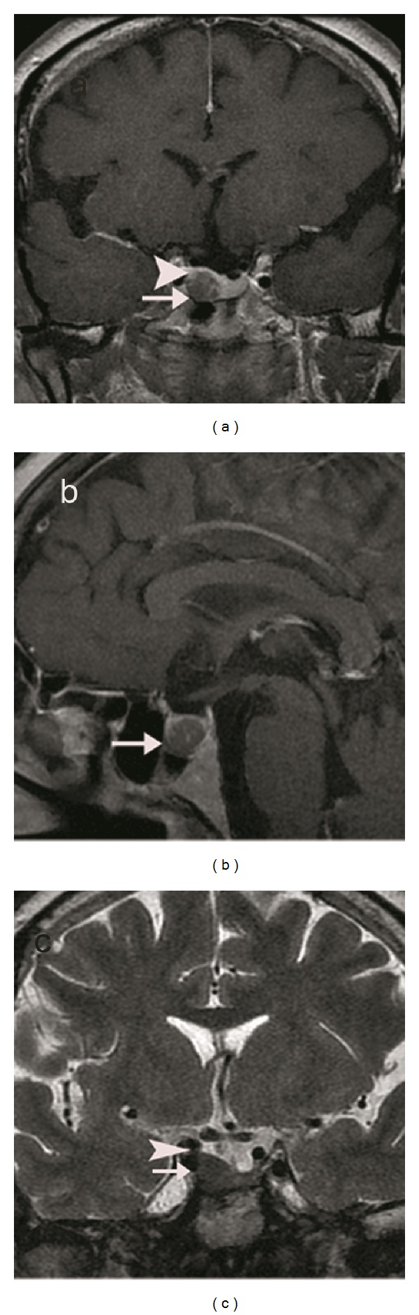
Magnetic resonance imaging is the most important diagnostic method in the investigation of the pituitary lesions. Our aim is to determine whether T2-weighted coronal images may be helpful in the evaluation of the pituitary gland with suspected pituitary adenomas. One hundred and sixty-seven patients were examined prospectively with T2-weighted coronal and T1-weighted coronal images enhanced with intravenous contrast material. The images were evaluated for the presence, the size, the location, and the ancillary signs including sellar floor erosion or ballooning, infindibulary deviation, convexity of the superior border of the gland, diffuse enlargement of the gland, and the invasion of the cavenous sinuses on both images. In forty-six (28%) patients lesions were revealed on both sequences. In twenty-one (12%) patients the lesions that were revealed on the T1-weighted images were not detected on the T2-weighted images. Positive predictive value, negative predictive value, sensitivity, specificity, and diagnostic accuracy rates of T2-weighted coronal images on the detection of the presence of lesions were 100%, 17.4%, 68.7%, 100%, and 87.4%, respectively. Both T2-weighted coronal and T1-weighted coronal images enhanced with intravenous gadolinium-based contrast material are important in the diagnosis of pituitary adenomas. T2-weighted coronal images could be used as a screening tool for the primary evaluation of the pituitary gland.

 求助内容:
求助内容: 应助结果提醒方式:


