Diagnostic Accuracy of Contrast-Enhanced FLAIR Magnetic Resonance Imaging in Diagnosis of Meningitis Correlated with CSF Analysis.
引用次数: 40
Abstract
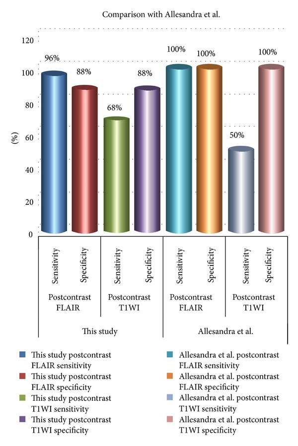
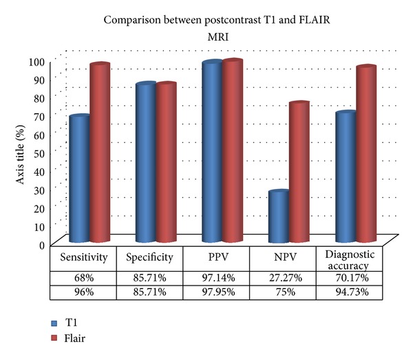
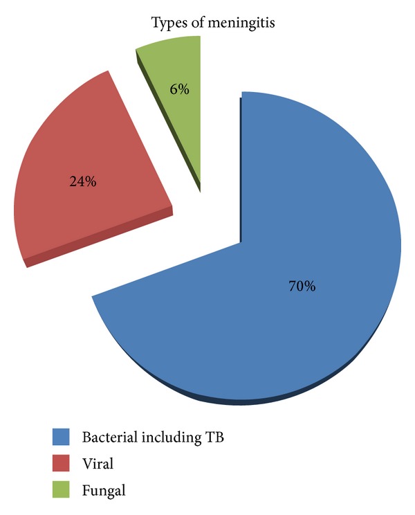
Purpose. To determine the diagnostic accuracy of contrast enhanced FLAIR sequence of MRI brain in the diagnosis of meningitis. Subjects and Methods. A prospective study of 57 patients with signs and symptoms of meningitis, referred to the radiology department for MRI examination. Out of these, there were 30 males and 27 females. They underwent MRI brain with contrast including postcontrast T1W and FLAIR sequences. Cerebrospinal fluid (CSF) analysis obtained by lumbar puncture after MRI was considered the “reference standard” against which MRI findings were compared. Results. Of 57 patients, 50 were diagnosed as having meningitis on subsequent CSF analysis. Out of these 50, 49 were positive on postcontrast FLAIR images and 34 were positive on postcontrast T1W images. One patient was labeled false positive as CSF analysis showed malignant cells (leptomeningeal carcinomatosis). In the diagnosis of meningitis, the sensitivity of postcontrast FLAIR sequence was 96% and specificity 85.71%, whereas the sensitivity of postcontrast T1W sequence was 68% and specificity 85.71%. Conclusion. Contrast-enhanced FLAIR sequence is more sensitive and specific than contrast-enhanced T1W sequence in the diagnosis of meningitis. It should be routinely used in suspected cases of meningitis.
对比增强FLAIR磁共振诊断脑膜炎的准确性与脑脊液分析的相关性。
目的。目的探讨脑MRI增强FLAIR序列对脑膜炎的诊断准确性。研究对象和方法。对57例有脑膜炎体征和症状的患者进行前瞻性研究,并转介到放射科进行MRI检查。其中,有30名男性和27名女性。他们接受了MRI脑造影,包括造影后的T1W和FLAIR序列。MRI后腰椎穿刺获得的脑脊液(CSF)分析被认为是与MRI结果进行比较的“参考标准”。结果。57例患者中,50例经脑脊液分析诊断为脑膜炎。在这50人中,49人在FLAIR图像上呈阳性,34人在T1W图像上呈阳性。一名患者被标记为假阳性,因为脑脊液分析显示恶性细胞(小脑脊膜癌)。FLAIR序列对脑膜炎的诊断敏感性为96%,特异性为85.71%,T1W序列对脑膜炎的诊断敏感性为68%,特异性为85.71%。结论。对比增强FLAIR序列比对比增强T1W序列在脑膜炎诊断中的敏感性和特异性更高。对于疑似脑膜炎病例,应常规使用。
本文章由计算机程序翻译,如有差异,请以英文原文为准。
求助全文
约1分钟内获得全文
求助全文

 求助内容:
求助内容: 应助结果提醒方式:


