Everli P S Gonçalves Gomes, Carlos Eduardo Rochitte, Clerio F Azevedo, Pedro A Lemos, Paulo Sampaio Gutierrez, Luiz Antonio M César
{"title":"Ex-vivo Assessment of Coronary Artery Atherosclerosis by Magnetic Resonance Imaging: Correlation with Histopathology.","authors":"Everli P S Gonçalves Gomes, Carlos Eduardo Rochitte, Clerio F Azevedo, Pedro A Lemos, Paulo Sampaio Gutierrez, Luiz Antonio M César","doi":"10.2174/1874192401408010026","DOIUrl":null,"url":null,"abstract":"<p><strong>Introduction: </strong>In recent years, high-resolution magnetic resonance imaging (MRI) has emerged as a very promising technique for studying atherosclerotic disease in humans.</p><p><strong>Aim: </strong>In the present study we sought to determine whether MRI allowed for the morphological characterization of the coronary vessel wall and atherosclerotic plaques using histopathological assessment as the reference standard.</p><p><strong>Methods: </strong>The study population consisted of 13 patients who died of acute myocardial infarction and underwent autopsy. The proximal portions of the coronary arteries were excised and were evaluated both by MRI and by histopathology. For each arterial segment, the following parameters were calculated through manual planimetry: 1. total vessel area (TVA); 2. luminal area (LA) and 3. plaque area (PA).</p><p><strong>Results: </strong>A total of 207 coronary artery cross-sections were found to be suitable for analysis by both MRI and histopathology and were included in the final analyses. Both methods demonstrated moderate to good agreement for the quantification of TVA (mean difference = 2.4±2.4 mm(2), 95‰ limits of agreement from -2.4 to +7.2 mm(2); CCC = 0.69, 95‰ CI from 0.63 to 0.75), LA (mean difference = 0.0±1.7 mm(2), 95‰ limits of agreement from -3.3 to + 3.3 mm(2); CCC = 0.84, 95‰ CI from 0.80 to 0.88) and PA (mean difference = 2.4±2.4 mm(2), 95‰ limits of agreement from -2.3 to + 7.1 mm(2); CCC = 0.64, 95‰ CI from 0.58 to 0.71).</p><p><strong>Conclusion: </strong>In this ex vivo experimental model we demonstrated good agreement between coronary artery morphometrical measurements obtained by high-resolution MRI and by histopathology.</p>","PeriodicalId":504447,"journal":{"name":"The Open Cardiovascular Medicine Journal","volume":"8 ","pages":"26-34"},"PeriodicalIF":0.0000,"publicationDate":"2014-04-04","publicationTypes":"Journal Article","fieldsOfStudy":null,"isOpenAccess":false,"openAccessPdf":"https://ftp.ncbi.nlm.nih.gov/pub/pmc/oa_pdf/48/8f/TOCMJ-8-26.PMC4021207.pdf","citationCount":"1","resultStr":null,"platform":"Semanticscholar","paperid":null,"PeriodicalName":"The Open Cardiovascular Medicine Journal","FirstCategoryId":"1085","ListUrlMain":"https://doi.org/10.2174/1874192401408010026","RegionNum":0,"RegionCategory":null,"ArticlePicture":[],"TitleCN":null,"AbstractTextCN":null,"PMCID":null,"EPubDate":"2014/1/1 0:00:00","PubModel":"eCollection","JCR":"","JCRName":"","Score":null,"Total":0}
引用次数: 1
Abstract
Introduction: In recent years, high-resolution magnetic resonance imaging (MRI) has emerged as a very promising technique for studying atherosclerotic disease in humans.
Aim: In the present study we sought to determine whether MRI allowed for the morphological characterization of the coronary vessel wall and atherosclerotic plaques using histopathological assessment as the reference standard.
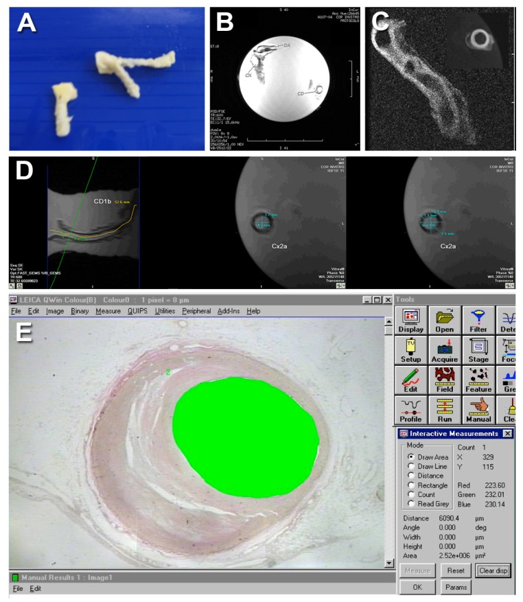
Methods: The study population consisted of 13 patients who died of acute myocardial infarction and underwent autopsy. The proximal portions of the coronary arteries were excised and were evaluated both by MRI and by histopathology. For each arterial segment, the following parameters were calculated through manual planimetry: 1. total vessel area (TVA); 2. luminal area (LA) and 3. plaque area (PA).
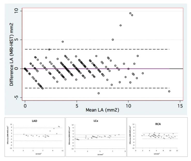
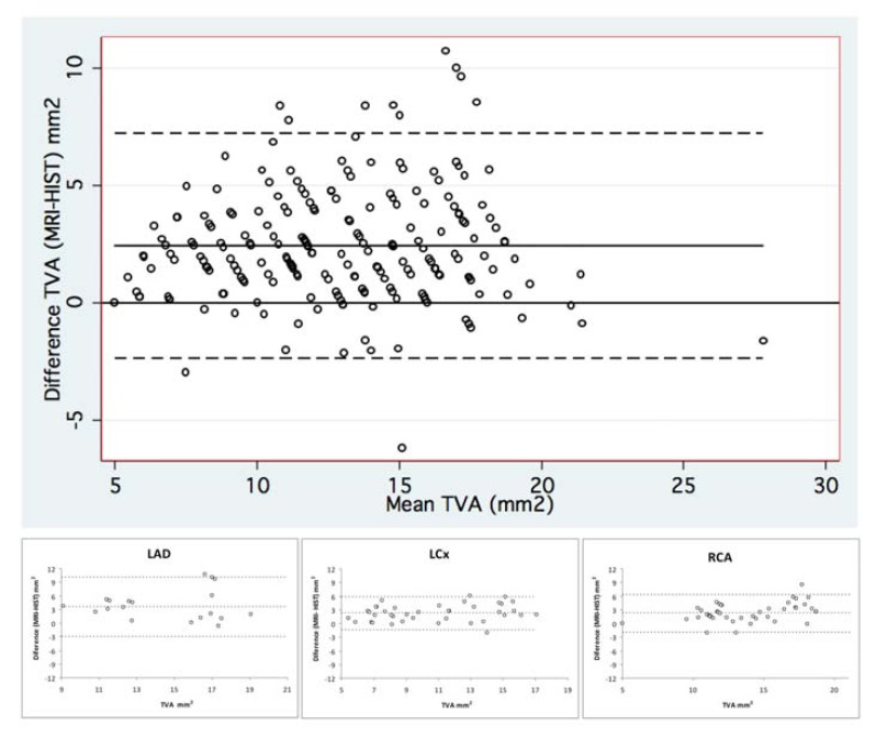
Results: A total of 207 coronary artery cross-sections were found to be suitable for analysis by both MRI and histopathology and were included in the final analyses. Both methods demonstrated moderate to good agreement for the quantification of TVA (mean difference = 2.4±2.4 mm(2), 95‰ limits of agreement from -2.4 to +7.2 mm(2); CCC = 0.69, 95‰ CI from 0.63 to 0.75), LA (mean difference = 0.0±1.7 mm(2), 95‰ limits of agreement from -3.3 to + 3.3 mm(2); CCC = 0.84, 95‰ CI from 0.80 to 0.88) and PA (mean difference = 2.4±2.4 mm(2), 95‰ limits of agreement from -2.3 to + 7.1 mm(2); CCC = 0.64, 95‰ CI from 0.58 to 0.71).
Conclusion: In this ex vivo experimental model we demonstrated good agreement between coronary artery morphometrical measurements obtained by high-resolution MRI and by histopathology.

 求助内容:
求助内容: 应助结果提醒方式:


