Nazir A Khan, Mudasir Ul Islam, Ayaz Ur Rehman, Shakeel Ahmad
{"title":"Pseudocyst of pinna and its treatment with surgical deroofing: an experience at tertiary hospitals.","authors":"Nazir A Khan, Mudasir Ul Islam, Ayaz Ur Rehman, Shakeel Ahmad","doi":"10.4103/2006-8808.128728","DOIUrl":null,"url":null,"abstract":"<p><strong>Introduction: </strong>Pseudocyst of pinna is an uncommon condition hardly encountered in routine ENT practice. The involvement is usually seen in scaphoid, triangular fossa, and antihelix. Medical treatment is ineffective. Various treatments are suggested in the literature. The aims of the paper were to study the clinical characteristic of patients with pseudocysts and to share our experience with surgical deroofing and buttoning as a definitive treatment.</p><p><strong>Materials and methods: </strong>Twenty-six patients were diagnosed with pseudocyst of the auricle between April 2011 and 2013 in two medical college hospitals. Clinical characteristics were noted. All patients underwent incision and drainage with removal of anterior cartilage leaflet followed by buttoning for 12 days.</p><p><strong>Results and observations: </strong>Out of 26 patients, only two were females. Involvement of left side was seen more than right one. None had bilateral involvement. Adults in the age group of 31-40 were commonly affected. Most common site of involvement was scaphoid and triangular fossa. The success rate with primary I and D and buttoning was 96%.</p><p><strong>Conclusions: </strong>Pseudocyst of the pinna is a benign condition of unknown etiology affecting the pinna, commonly encountered in middle-aged men. Many modalities of treatment have been recommended in the literature with varied recurrence and failure rates. The best form of treatment with minimum recurrence is incision and drainage with removal of anterior cartilage leaflet with buttoning.</p>","PeriodicalId":89430,"journal":{"name":"Journal of surgical technique and case report","volume":"5 2","pages":"72-7"},"PeriodicalIF":0.0000,"publicationDate":"2013-07-01","publicationTypes":"Journal Article","fieldsOfStudy":null,"isOpenAccess":false,"openAccessPdf":"https://sci-hub-pdf.com/10.4103/2006-8808.128728","citationCount":"17","resultStr":null,"platform":"Semanticscholar","paperid":null,"PeriodicalName":"Journal of surgical technique and case report","FirstCategoryId":"1085","ListUrlMain":"https://doi.org/10.4103/2006-8808.128728","RegionNum":0,"RegionCategory":null,"ArticlePicture":[],"TitleCN":null,"AbstractTextCN":null,"PMCID":null,"EPubDate":"","PubModel":"","JCR":"","JCRName":"","Score":null,"Total":0}
引用次数: 17
Abstract
Introduction: Pseudocyst of pinna is an uncommon condition hardly encountered in routine ENT practice. The involvement is usually seen in scaphoid, triangular fossa, and antihelix. Medical treatment is ineffective. Various treatments are suggested in the literature. The aims of the paper were to study the clinical characteristic of patients with pseudocysts and to share our experience with surgical deroofing and buttoning as a definitive treatment.
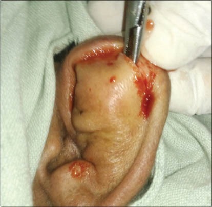
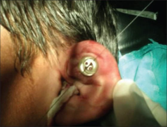
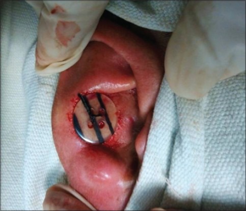
Materials and methods: Twenty-six patients were diagnosed with pseudocyst of the auricle between April 2011 and 2013 in two medical college hospitals. Clinical characteristics were noted. All patients underwent incision and drainage with removal of anterior cartilage leaflet followed by buttoning for 12 days.
Results and observations: Out of 26 patients, only two were females. Involvement of left side was seen more than right one. None had bilateral involvement. Adults in the age group of 31-40 were commonly affected. Most common site of involvement was scaphoid and triangular fossa. The success rate with primary I and D and buttoning was 96%.
Conclusions: Pseudocyst of the pinna is a benign condition of unknown etiology affecting the pinna, commonly encountered in middle-aged men. Many modalities of treatment have been recommended in the literature with varied recurrence and failure rates. The best form of treatment with minimum recurrence is incision and drainage with removal of anterior cartilage leaflet with buttoning.

 求助内容:
求助内容: 应助结果提醒方式:


