{"title":"Characterization of the liver-macrophages isolated from a mixed primary culture of neonatal swine hepatocytes","authors":"Hiroshi Kitani , Miyako Yoshioka , Takato Takenouchi , Mitsuru Sato , Noriko Yamanaka","doi":"10.1016/j.rinim.2014.01.001","DOIUrl":null,"url":null,"abstract":"<div><p>We recently developed a novel procedure to obtain liver-macrophages in sufficient number and purity using a mixed primary culture of rat and bovine hepatocytes. In this study, we aim to apply this method to the neonatal swine liver. Swine parenchymal hepatocytes were isolated by a two-step collagenase perfusion method and cultured in T75 culture flasks. Similar to the rat and bovine cells, the swine hepatocytes retained an epithelial cell morphology for only a few days and progressively changed into fibroblastic cells. After 5–13 days of culture, macrophage-like cells actively proliferated on the mixed fibroblastic cell sheet. Gentle shaking of the culture flask followed by the transfer and brief incubation of the culture supernatant resulted in a quick and selective adhesion of macrophage-like cells to a plastic dish surface. After rinsing dishes with saline, the attached macrophage-like cells were collected at a yield of 10<sup>6</sup> cells per T75 culture flask at 2–3 day intervals for more than 3 weeks. The isolated cells displayed a typical macrophage morphology and were strongly positive for macrophage markers, such as CD172a, Iba-1 and KT022, but negative for cytokeratin, desmin and α-smooth muscle actin, indicating a highly purified macrophage population. The isolated cells exhibited phagocytosis of polystyrene microbeads and a release of inflammatory cytokines upon lipopolysaccharide stimulation. This shaking and attachment method is applicable to the swine liver and provides a sufficient number of macrophages without any need of complex laboratory equipments.</p></div>","PeriodicalId":89845,"journal":{"name":"Results in immunology","volume":"4 ","pages":"Pages 1-7"},"PeriodicalIF":0.0000,"publicationDate":"2014-01-01","publicationTypes":"Journal Article","fieldsOfStudy":null,"isOpenAccess":false,"openAccessPdf":"https://sci-hub-pdf.com/10.1016/j.rinim.2014.01.001","citationCount":"13","resultStr":null,"platform":"Semanticscholar","paperid":null,"PeriodicalName":"Results in immunology","FirstCategoryId":"1085","ListUrlMain":"https://www.sciencedirect.com/science/article/pii/S2211283914000021","RegionNum":0,"RegionCategory":null,"ArticlePicture":[],"TitleCN":null,"AbstractTextCN":null,"PMCID":null,"EPubDate":"","PubModel":"","JCR":"","JCRName":"","Score":null,"Total":0}
引用次数: 13
Abstract
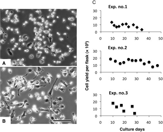
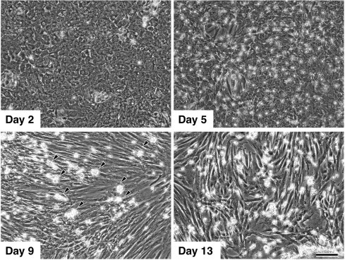
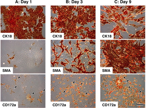
We recently developed a novel procedure to obtain liver-macrophages in sufficient number and purity using a mixed primary culture of rat and bovine hepatocytes. In this study, we aim to apply this method to the neonatal swine liver. Swine parenchymal hepatocytes were isolated by a two-step collagenase perfusion method and cultured in T75 culture flasks. Similar to the rat and bovine cells, the swine hepatocytes retained an epithelial cell morphology for only a few days and progressively changed into fibroblastic cells. After 5–13 days of culture, macrophage-like cells actively proliferated on the mixed fibroblastic cell sheet. Gentle shaking of the culture flask followed by the transfer and brief incubation of the culture supernatant resulted in a quick and selective adhesion of macrophage-like cells to a plastic dish surface. After rinsing dishes with saline, the attached macrophage-like cells were collected at a yield of 106 cells per T75 culture flask at 2–3 day intervals for more than 3 weeks. The isolated cells displayed a typical macrophage morphology and were strongly positive for macrophage markers, such as CD172a, Iba-1 and KT022, but negative for cytokeratin, desmin and α-smooth muscle actin, indicating a highly purified macrophage population. The isolated cells exhibited phagocytosis of polystyrene microbeads and a release of inflammatory cytokines upon lipopolysaccharide stimulation. This shaking and attachment method is applicable to the swine liver and provides a sufficient number of macrophages without any need of complex laboratory equipments.

 求助内容:
求助内容: 应助结果提醒方式:


