Chih-Wen Ni, Sandeep Kumar, Casey J Ankeny, Hanjoong Jo
{"title":"Development of immortalized mouse aortic endothelial cell lines.","authors":"Chih-Wen Ni, Sandeep Kumar, Casey J Ankeny, Hanjoong Jo","doi":"10.1186/2045-824X-6-7","DOIUrl":null,"url":null,"abstract":"<p><strong>Background: </strong>The understanding of endothelial cell biology has been facilitated by the availability of primary endothelial cell cultures from a variety of sites and species; however, the isolation and maintenance of primary mouse aortic endothelial cells (MAECs) remain a formidable challenge. Culturing MAECs is difficult as they are prone to phenotypic drift during culture. Therefore, there is a need to have a dependable in vitro culture system, wherein the primary endothelial cells retain their properties and phenotypes.</p><p><strong>Methods: </strong>Here, we developed an effective method to prepare immortalized MAEC (iMAEC) lines. Primary MAECs, initially isolated from aortic explants, were immortalized using a retrovirus expressing polyoma middle T-antigen. Immortalized cells were then incubated with DiI-acetylated-low density lipoprotein and sorted via flow cytometry to isolate iMAECs.</p><p><strong>Results: </strong>iMAECs expressed common markers of endothelial cells, including PECAM1, eNOS, VE-cadherin, and von Willebrand Factor. iMAECs aligned in the direction of imposed laminar shear and retained the ability to form tubes. Using this method, we have generated iMAEC lines from wild-type and various genetically modified mice such as p47phox-/-, eNOS-/-, and caveolin-1-/-.</p><p><strong>Conclusion: </strong>In summary, generation of iMAEC lines from various genetically modified mouse lines provides an invaluable tool to study vascular biology and pathophysiology.</p>","PeriodicalId":23948,"journal":{"name":"Vascular Cell","volume":"6 1","pages":"7"},"PeriodicalIF":0.0000,"publicationDate":"2014-04-01","publicationTypes":"Journal Article","fieldsOfStudy":null,"isOpenAccess":false,"openAccessPdf":"https://sci-hub-pdf.com/10.1186/2045-824X-6-7","citationCount":"33","resultStr":null,"platform":"Semanticscholar","paperid":null,"PeriodicalName":"Vascular Cell","FirstCategoryId":"1085","ListUrlMain":"https://doi.org/10.1186/2045-824X-6-7","RegionNum":0,"RegionCategory":null,"ArticlePicture":[],"TitleCN":null,"AbstractTextCN":null,"PMCID":null,"EPubDate":"","PubModel":"","JCR":"Q4","JCRName":"Neuroscience","Score":null,"Total":0}
引用次数: 33
Abstract
Background: The understanding of endothelial cell biology has been facilitated by the availability of primary endothelial cell cultures from a variety of sites and species; however, the isolation and maintenance of primary mouse aortic endothelial cells (MAECs) remain a formidable challenge. Culturing MAECs is difficult as they are prone to phenotypic drift during culture. Therefore, there is a need to have a dependable in vitro culture system, wherein the primary endothelial cells retain their properties and phenotypes.
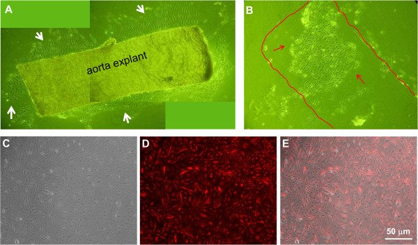
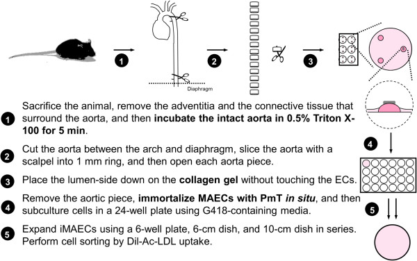
Methods: Here, we developed an effective method to prepare immortalized MAEC (iMAEC) lines. Primary MAECs, initially isolated from aortic explants, were immortalized using a retrovirus expressing polyoma middle T-antigen. Immortalized cells were then incubated with DiI-acetylated-low density lipoprotein and sorted via flow cytometry to isolate iMAECs.
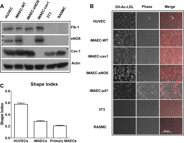
Results: iMAECs expressed common markers of endothelial cells, including PECAM1, eNOS, VE-cadherin, and von Willebrand Factor. iMAECs aligned in the direction of imposed laminar shear and retained the ability to form tubes. Using this method, we have generated iMAEC lines from wild-type and various genetically modified mice such as p47phox-/-, eNOS-/-, and caveolin-1-/-.
Conclusion: In summary, generation of iMAEC lines from various genetically modified mouse lines provides an invaluable tool to study vascular biology and pathophysiology.

 求助内容:
求助内容: 应助结果提醒方式:


