{"title":"A case of giant osteoma developed from the mastoid cortical bone.","authors":"Sung Joon Park, Young Ho Kim","doi":"10.7874/kja.2012.16.2.95","DOIUrl":null,"url":null,"abstract":"<p><p>An osteoma of the temporal bone is a rare benign tumor. External auditory canal is the most common site of osteomas arising from temporal bone, and mastoid osteoma is very rare. A case of a 42-year-old female with a huge osteoma which developed from mastoid cortical bone is presented and the review of the temporal bone osteomas is discussed. The patient showed a huge and hard mass in the right mastoid area growing over a 20-year period. A temporal bone computed tomography scan demonstrated 2.3×2.3×4.3 cm sized bony tumor on surface of the right mastoid and squama. The resection of whole bony tumor with mastoid cortical bone was performed using retroauricular approach. Pathologic evaluation revealed the osteoma. The huge osteoma in the mastoid area may induce a cosmetic deformity. Early diagnosis and surgical removal of the osteoma may ensure an easy and complete treatment. The total resection of bony tumor including mastoid cortical bone is recommended to avoid recurrence. </p>","PeriodicalId":90252,"journal":{"name":"Korean journal of audiology","volume":"16 2","pages":"95-8"},"PeriodicalIF":0.0000,"publicationDate":"2012-09-01","publicationTypes":"Journal Article","fieldsOfStudy":null,"isOpenAccess":false,"openAccessPdf":"https://ftp.ncbi.nlm.nih.gov/pub/pmc/oa_pdf/18/8a/kja-16-95.PMC3936565.pdf","citationCount":"21","resultStr":null,"platform":"Semanticscholar","paperid":null,"PeriodicalName":"Korean journal of audiology","FirstCategoryId":"1085","ListUrlMain":"https://doi.org/10.7874/kja.2012.16.2.95","RegionNum":0,"RegionCategory":null,"ArticlePicture":[],"TitleCN":null,"AbstractTextCN":null,"PMCID":null,"EPubDate":"2012/9/20 0:00:00","PubModel":"Epub","JCR":"","JCRName":"","Score":null,"Total":0}
引用次数: 21
Abstract
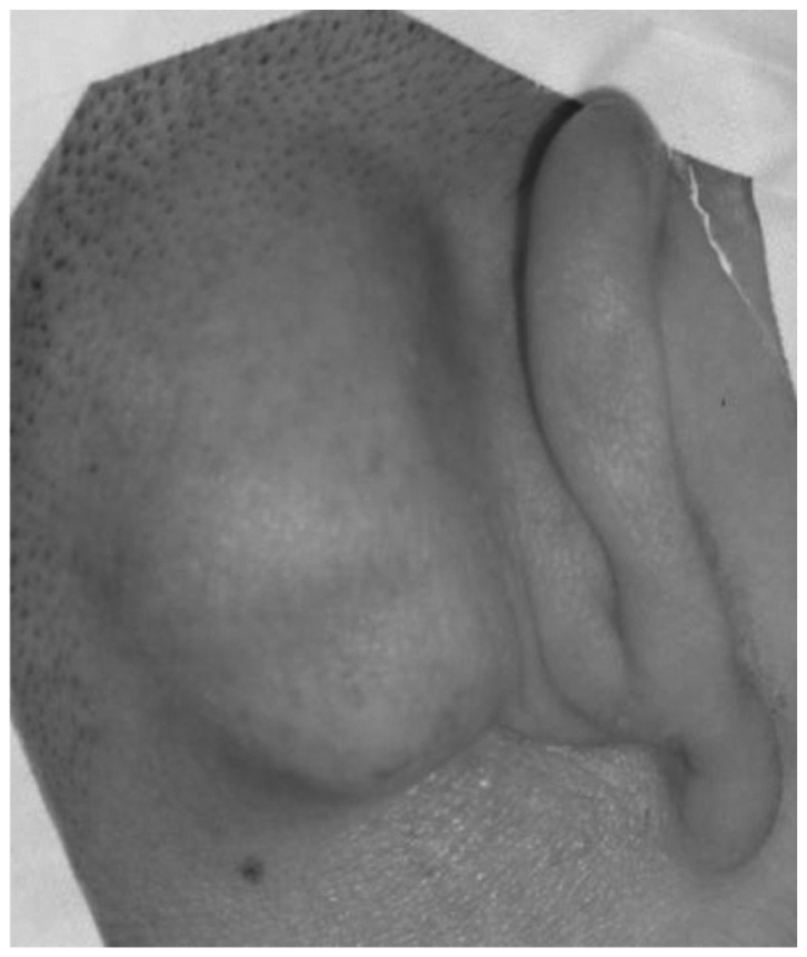
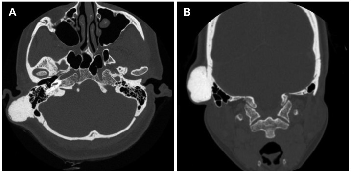
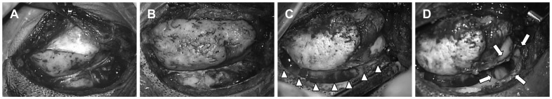
An osteoma of the temporal bone is a rare benign tumor. External auditory canal is the most common site of osteomas arising from temporal bone, and mastoid osteoma is very rare. A case of a 42-year-old female with a huge osteoma which developed from mastoid cortical bone is presented and the review of the temporal bone osteomas is discussed. The patient showed a huge and hard mass in the right mastoid area growing over a 20-year period. A temporal bone computed tomography scan demonstrated 2.3×2.3×4.3 cm sized bony tumor on surface of the right mastoid and squama. The resection of whole bony tumor with mastoid cortical bone was performed using retroauricular approach. Pathologic evaluation revealed the osteoma. The huge osteoma in the mastoid area may induce a cosmetic deformity. Early diagnosis and surgical removal of the osteoma may ensure an easy and complete treatment. The total resection of bony tumor including mastoid cortical bone is recommended to avoid recurrence.

 求助内容:
求助内容: 应助结果提醒方式:


