Hyun Jung Min, Chul Won Park, Jin Hyeok Jeong, Seok Hyun Cho, Kyung Rae Kim, Seung Hwan Lee
{"title":"Comparative analysis of the expression of involucrin, filaggrin and cytokeratin 4, 10, 16 in cholesteatoma.","authors":"Hyun Jung Min, Chul Won Park, Jin Hyeok Jeong, Seok Hyun Cho, Kyung Rae Kim, Seung Hwan Lee","doi":"10.7874/kja.2012.16.3.124","DOIUrl":null,"url":null,"abstract":"<p><strong>Background and objectives: </strong>The aim of this study is to determine whether the hyperproliferative and hyperkeratotic characters of cholesteatoma are associated with differentiation of keratinocytes in cholesteatoma by examining the localization of marker proteins, such as involucrin, filaggrin, and cytokeratins.</p><p><strong>Materials and methods: </strong>Immunohistochemical study was carried out in 30 cholesteatoma tissues and 10 retroauricular skins to examine the expression of involucrin, filaggrin, cytokeratin 4, 10 and 16. The staining results were graded as negative, weakly positive (<10%), moderately positive (10-70%), and strongly positive (>70%).</p><p><strong>Results: </strong>Involucrin was strongly expressed in upper spinous, granular, and corneal layer of cholesteatoma. Filaggrin was strongly expressed in granular and corneal layer of cholesteatoma. Cytokeratin 4 was expressed in basal layer of retroauricular skin, but occasionally expressed in suprabasal layer of cholesteatoma. Cytokeratin 10 was homogenously expressed in all suprabasal layer of retroauricular skin, whereas pattern of shift to surface layer was showed in cholesteatoma. Cytokeratin 16 was moderately expressed at suprabasal layer in cholesteatoma.</p><p><strong>Conclusions: </strong>It can be suggested that early differentiation of suprabasal layer may lead to hyperdifferentiation and hyperkeratosis. Different expression of cytokeratins possibly indicates the altered differentiation of cholesteatoma.</p>","PeriodicalId":90252,"journal":{"name":"Korean journal of audiology","volume":"16 3","pages":"124-9"},"PeriodicalIF":0.0000,"publicationDate":"2012-12-01","publicationTypes":"Journal Article","fieldsOfStudy":null,"isOpenAccess":false,"openAccessPdf":"https://ftp.ncbi.nlm.nih.gov/pub/pmc/oa_pdf/66/10/kja-16-124.PMC3936667.pdf","citationCount":"6","resultStr":null,"platform":"Semanticscholar","paperid":null,"PeriodicalName":"Korean journal of audiology","FirstCategoryId":"1085","ListUrlMain":"https://doi.org/10.7874/kja.2012.16.3.124","RegionNum":0,"RegionCategory":null,"ArticlePicture":[],"TitleCN":null,"AbstractTextCN":null,"PMCID":null,"EPubDate":"2012/12/18 0:00:00","PubModel":"Epub","JCR":"","JCRName":"","Score":null,"Total":0}
引用次数: 6
Abstract
Background and objectives: The aim of this study is to determine whether the hyperproliferative and hyperkeratotic characters of cholesteatoma are associated with differentiation of keratinocytes in cholesteatoma by examining the localization of marker proteins, such as involucrin, filaggrin, and cytokeratins.
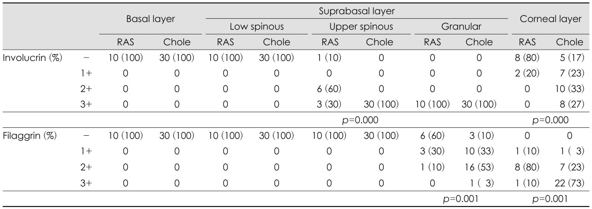
Materials and methods: Immunohistochemical study was carried out in 30 cholesteatoma tissues and 10 retroauricular skins to examine the expression of involucrin, filaggrin, cytokeratin 4, 10 and 16. The staining results were graded as negative, weakly positive (<10%), moderately positive (10-70%), and strongly positive (>70%).
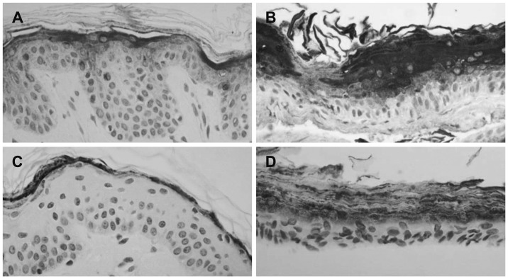
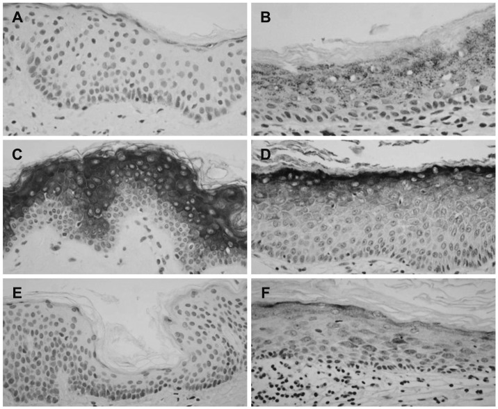
Results: Involucrin was strongly expressed in upper spinous, granular, and corneal layer of cholesteatoma. Filaggrin was strongly expressed in granular and corneal layer of cholesteatoma. Cytokeratin 4 was expressed in basal layer of retroauricular skin, but occasionally expressed in suprabasal layer of cholesteatoma. Cytokeratin 10 was homogenously expressed in all suprabasal layer of retroauricular skin, whereas pattern of shift to surface layer was showed in cholesteatoma. Cytokeratin 16 was moderately expressed at suprabasal layer in cholesteatoma.
Conclusions: It can be suggested that early differentiation of suprabasal layer may lead to hyperdifferentiation and hyperkeratosis. Different expression of cytokeratins possibly indicates the altered differentiation of cholesteatoma.

 求助内容:
求助内容: 应助结果提醒方式:


