Byoung Soo Shim, Byung Chul Kang, Chang-Hee Kim, Tae Su Kim, Hong Ju Park
{"title":"Superior canal dehiscence patients have smaller mastoid volume than age- and sex-matched otosclerosis and temporal bone fracture patients.","authors":"Byoung Soo Shim, Byung Chul Kang, Chang-Hee Kim, Tae Su Kim, Hong Ju Park","doi":"10.7874/kja.2012.16.3.120","DOIUrl":null,"url":null,"abstract":"<p><strong>Background and objectives: </strong>The purpose of the study was to compare the mastoid air-cell volume of the patients with superior semicircular canal dehiscence syndrome (SCDS) and that of the control patients with otosclerosis and temporal bone (TB) fracture.</p><p><strong>Subjects and methods: </strong>Ten patients with SCDS were enrolled and 10 patients with bilateral otosclerosis and TB fracture were selected as control groups by age and sex matching. To measure the mastoid air-cell volume, 3D reconstruction software was used.</p><p><strong>Results: </strong>In 10 patients with SCDS, the mean age was 44.5 years, ranging from 16 to 79 years (M : F=4 : 6). Mean mastoid air-cell volume in the SCDS side was 3319.9 mm(3), whereas 4177.2 mm(3) in the normal side (p=0.022). Mean mastoid air-cell volume in the right side of otosclerosis patients was 6594.3 mm(3) and it was not different from 6380.5 mm(3) in the left side (p=0.445). Mean mastoid air-cell volume in normal side of TB fracture was 6477.2 mm(3). The mastoid air-cell volume in the SCDS side was significantly smaller than that of otosclerosis and TB fracture patients (p=0.009, p=0.002, respectively). The mastoid air-cell volume in the normal side of SCDS was significantly smaller than that of TB fracture (p=0.019), but not significant with that of otosclerosis (p=0.063).</p><p><strong>Conclusions: </strong>Our findings revealed that the mastoid air-cell volume in the SCDS side was significantly smaller than control group, which suggest that the decreased mastoid pneumatization is closely related to the generation of SCDS.</p>","PeriodicalId":90252,"journal":{"name":"Korean journal of audiology","volume":"16 3","pages":"120-3"},"PeriodicalIF":0.0000,"publicationDate":"2012-12-01","publicationTypes":"Journal Article","fieldsOfStudy":null,"isOpenAccess":false,"openAccessPdf":"https://ftp.ncbi.nlm.nih.gov/pub/pmc/oa_pdf/7f/aa/kja-16-120.PMC3936659.pdf","citationCount":"14","resultStr":null,"platform":"Semanticscholar","paperid":null,"PeriodicalName":"Korean journal of audiology","FirstCategoryId":"1085","ListUrlMain":"https://doi.org/10.7874/kja.2012.16.3.120","RegionNum":0,"RegionCategory":null,"ArticlePicture":[],"TitleCN":null,"AbstractTextCN":null,"PMCID":null,"EPubDate":"2012/12/18 0:00:00","PubModel":"Epub","JCR":"","JCRName":"","Score":null,"Total":0}
引用次数: 14
Abstract
Background and objectives: The purpose of the study was to compare the mastoid air-cell volume of the patients with superior semicircular canal dehiscence syndrome (SCDS) and that of the control patients with otosclerosis and temporal bone (TB) fracture.
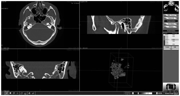
Subjects and methods: Ten patients with SCDS were enrolled and 10 patients with bilateral otosclerosis and TB fracture were selected as control groups by age and sex matching. To measure the mastoid air-cell volume, 3D reconstruction software was used.
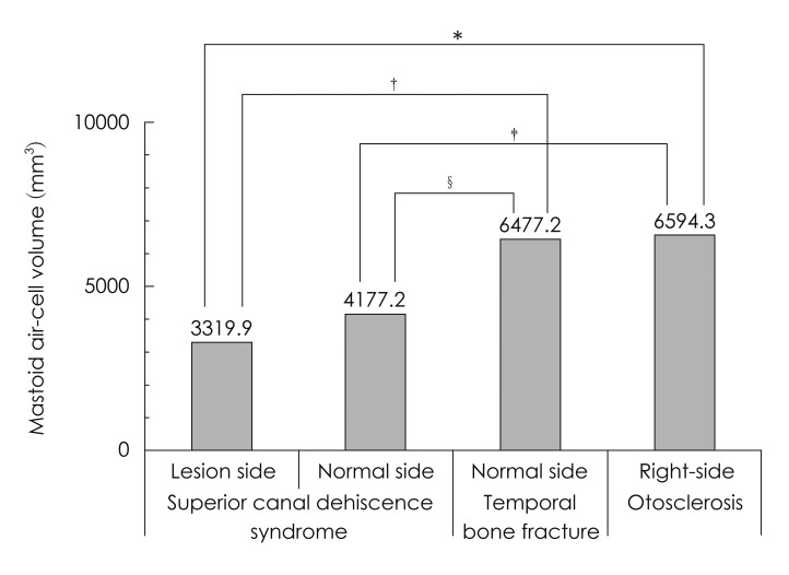
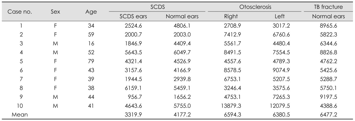
Results: In 10 patients with SCDS, the mean age was 44.5 years, ranging from 16 to 79 years (M : F=4 : 6). Mean mastoid air-cell volume in the SCDS side was 3319.9 mm(3), whereas 4177.2 mm(3) in the normal side (p=0.022). Mean mastoid air-cell volume in the right side of otosclerosis patients was 6594.3 mm(3) and it was not different from 6380.5 mm(3) in the left side (p=0.445). Mean mastoid air-cell volume in normal side of TB fracture was 6477.2 mm(3). The mastoid air-cell volume in the SCDS side was significantly smaller than that of otosclerosis and TB fracture patients (p=0.009, p=0.002, respectively). The mastoid air-cell volume in the normal side of SCDS was significantly smaller than that of TB fracture (p=0.019), but not significant with that of otosclerosis (p=0.063).
Conclusions: Our findings revealed that the mastoid air-cell volume in the SCDS side was significantly smaller than control group, which suggest that the decreased mastoid pneumatization is closely related to the generation of SCDS.

 求助内容:
求助内容: 应助结果提醒方式:


