Eimear Kennedy, Roya Hakimjavadi, Chris Greene, Ciaran J Mooney, Emma Fitzpatrick, Laura E Collins, Christine E Loscher, Shaunta Guha, David Morrow, Eileen M Redmond, Paul A Cahill
{"title":"Embryonic rat vascular smooth muscle cells revisited - a model for neonatal, neointimal SMC or differentiated vascular stem cells?","authors":"Eimear Kennedy, Roya Hakimjavadi, Chris Greene, Ciaran J Mooney, Emma Fitzpatrick, Laura E Collins, Christine E Loscher, Shaunta Guha, David Morrow, Eileen M Redmond, Paul A Cahill","doi":"10.1186/2045-824X-6-6","DOIUrl":null,"url":null,"abstract":"<p><strong>Background: </strong>The A10 and A7r5 cell lines derived from the thoracic aorta of embryonic rat are widely used as models of non-differentiated, neonatal and neointimal vascular smooth muscle cells in culture. The recent discovery of resident multipotent vascular stem cells within the vessel wall has necessitated the identity and origin of these vascular cells be revisited. In this context, we examined A10 and A7r5 cell lines to establish the similarities and differences between these cell lines and multipotent vascular stem cells isolated from adult rat aortas by determining their differentiation state, stem cell marker expression and their multipotency potential in vitro.</p><p><strong>Methods: </strong>Vascular smooth muscle cell differentiation markers (alpha-actin, myosin heavy chain, calponin) and stem cell marker expression (Sox10, Sox17 and S100β) were assessed using immunocytochemistry, confocal microscopy, FACS analysis and real-time quantitative PCR.</p><p><strong>Results: </strong>Both A10 and A7r5 expressed vascular smooth muscle differentiation, markers, smooth muscle alpha - actin, smooth muscle myosin heavy chain and calponin. In parallel analysis, multipotent vascular stem cells isolated from rat aortic explants were immunocytochemically myosin heavy chain negative but positive for the neural stem cell markers Sox10+, a neural crest marker, Sox17+ the endoderm marker, and the glia marker, S100β+. This multipotent vascular stem cell marker profile was detected in both embryonic vascular cell lines in addition to the adventitial progenitor stem cell marker, stem cell antigen-1, Sca1+. Serum deprivation resulted in a significant increase in stem cell and smooth muscle cell differentiation marker expression, when compared to serum treated cells. Both cell types exhibited weak multipotency following adipocyte inductive stimulation. Moreover, Notch signaling blockade following γ-secretase inhibition with DAPT enhanced the expression of both vascular smooth muscle and stem cell markers.</p><p><strong>Conclusions: </strong>We conclude that A10 and A7r5 cells share similar neural stem cell markers to both multipotent vascular stem cells and adventitial progenitors that are indicative of neointimal stem-derived smooth muscle cells. This may have important implications for their use in examining vascular contractile and proliferative phenotypes in vitro.</p>","PeriodicalId":23948,"journal":{"name":"Vascular Cell","volume":"6 1","pages":"6"},"PeriodicalIF":0.0000,"publicationDate":"2014-03-15","publicationTypes":"Journal Article","fieldsOfStudy":null,"isOpenAccess":false,"openAccessPdf":"https://sci-hub-pdf.com/10.1186/2045-824X-6-6","citationCount":"25","resultStr":null,"platform":"Semanticscholar","paperid":null,"PeriodicalName":"Vascular Cell","FirstCategoryId":"1085","ListUrlMain":"https://doi.org/10.1186/2045-824X-6-6","RegionNum":0,"RegionCategory":null,"ArticlePicture":[],"TitleCN":null,"AbstractTextCN":null,"PMCID":null,"EPubDate":"","PubModel":"","JCR":"Q4","JCRName":"Neuroscience","Score":null,"Total":0}
引用次数: 25
Abstract
Background: The A10 and A7r5 cell lines derived from the thoracic aorta of embryonic rat are widely used as models of non-differentiated, neonatal and neointimal vascular smooth muscle cells in culture. The recent discovery of resident multipotent vascular stem cells within the vessel wall has necessitated the identity and origin of these vascular cells be revisited. In this context, we examined A10 and A7r5 cell lines to establish the similarities and differences between these cell lines and multipotent vascular stem cells isolated from adult rat aortas by determining their differentiation state, stem cell marker expression and their multipotency potential in vitro.
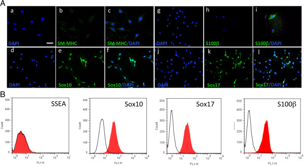
Methods: Vascular smooth muscle cell differentiation markers (alpha-actin, myosin heavy chain, calponin) and stem cell marker expression (Sox10, Sox17 and S100β) were assessed using immunocytochemistry, confocal microscopy, FACS analysis and real-time quantitative PCR.
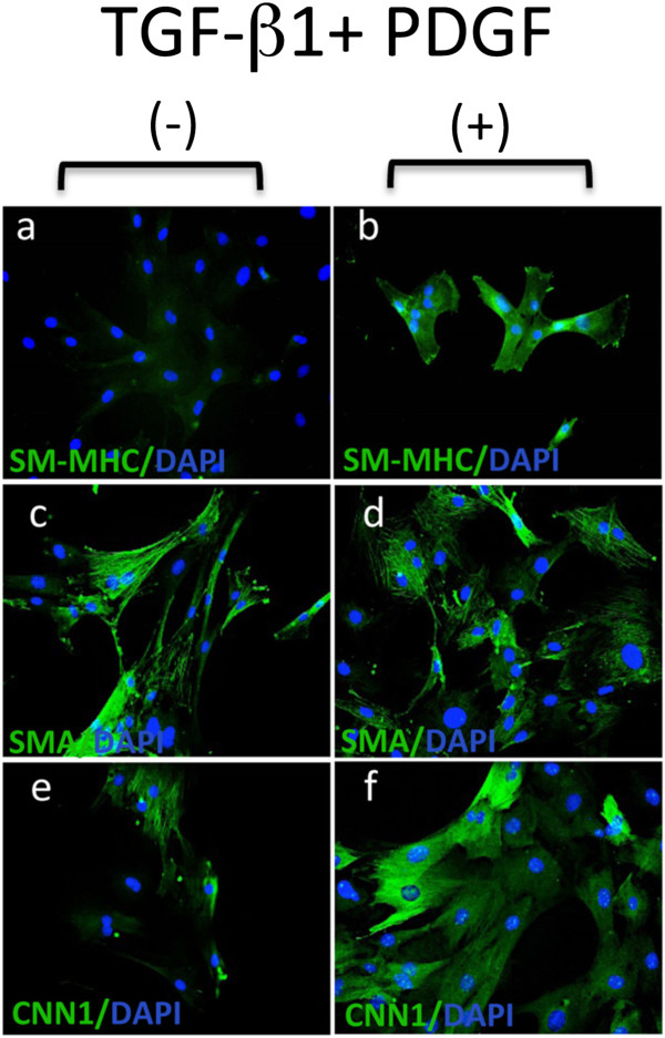
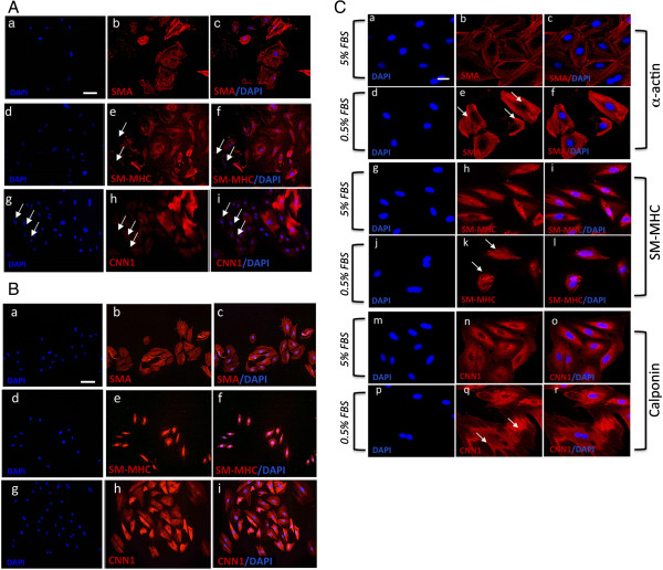
Results: Both A10 and A7r5 expressed vascular smooth muscle differentiation, markers, smooth muscle alpha - actin, smooth muscle myosin heavy chain and calponin. In parallel analysis, multipotent vascular stem cells isolated from rat aortic explants were immunocytochemically myosin heavy chain negative but positive for the neural stem cell markers Sox10+, a neural crest marker, Sox17+ the endoderm marker, and the glia marker, S100β+. This multipotent vascular stem cell marker profile was detected in both embryonic vascular cell lines in addition to the adventitial progenitor stem cell marker, stem cell antigen-1, Sca1+. Serum deprivation resulted in a significant increase in stem cell and smooth muscle cell differentiation marker expression, when compared to serum treated cells. Both cell types exhibited weak multipotency following adipocyte inductive stimulation. Moreover, Notch signaling blockade following γ-secretase inhibition with DAPT enhanced the expression of both vascular smooth muscle and stem cell markers.
Conclusions: We conclude that A10 and A7r5 cells share similar neural stem cell markers to both multipotent vascular stem cells and adventitial progenitors that are indicative of neointimal stem-derived smooth muscle cells. This may have important implications for their use in examining vascular contractile and proliferative phenotypes in vitro.

 求助内容:
求助内容: 应助结果提醒方式:


