Collision tumor of the kidney composed of clear cell carcinoma and collecting duct carcinoma: report of a case with unusual morphology and clinical follow-up.
{"title":"Collision tumor of the kidney composed of clear cell carcinoma and collecting duct carcinoma: report of a case with unusual morphology and clinical follow-up.","authors":"Rhonda Burch-Smith, Nizar M Tannir, Erika Resetkova, Pheroze Tamboli, Priya Rao","doi":"10.5732/cjc.013.10155","DOIUrl":null,"url":null,"abstract":"<p><p>We report the case of a 67-year-old female who presented with a large renal mass. Gross examination of the nephrectomy specimen demonstrated a 6-cm renal mass that invaded into the renal sinus and perinephric fat. Histologic examination revealed two distinct tumor types. The first type was a conventional (clear cell) renal cell carcinoma that was of low nuclear grade and comprised the minority of the overall tumor. The second type was a high-grade collecting duct carcinoma with glandular/tubular differentiation and composed the majority of the tumor. Immunohistochemical studies demonstrated distinctive patterns of the two tumor types, thus confirming two distinct lineages. Five months postoperatively, the patient developed metastasis to the lungs and right hilar lymph node region. A fine needle aspiration of a lung nodule demonstrated a metastatic, poorly differentiated carcinoma, similar to the collecting duct carcinoma component in the kidney. Collision tumors of the kidney are rare with fewer than 10 cases reported in the literature. Our report further expands the spectrum of this rare phenomenon. </p>","PeriodicalId":10034,"journal":{"name":"癌症","volume":"33 7","pages":"351-5"},"PeriodicalIF":0.0000,"publicationDate":"2014-07-01","publicationTypes":"Journal Article","fieldsOfStudy":null,"isOpenAccess":false,"openAccessPdf":"https://ftp.ncbi.nlm.nih.gov/pub/pmc/oa_pdf/e0/74/cjc-33-07-351.PMC4110468.pdf","citationCount":"13","resultStr":null,"platform":"Semanticscholar","paperid":null,"PeriodicalName":"癌症","FirstCategoryId":"3","ListUrlMain":"https://doi.org/10.5732/cjc.013.10155","RegionNum":0,"RegionCategory":null,"ArticlePicture":[],"TitleCN":null,"AbstractTextCN":null,"PMCID":null,"EPubDate":"2014/3/4 0:00:00","PubModel":"Epub","JCR":"Q","JCRName":"Medicine","Score":null,"Total":0}
引用次数: 13
Abstract
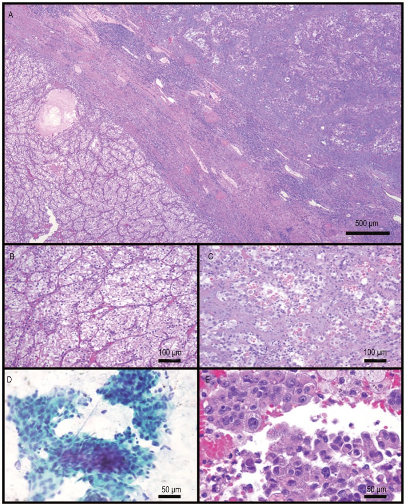
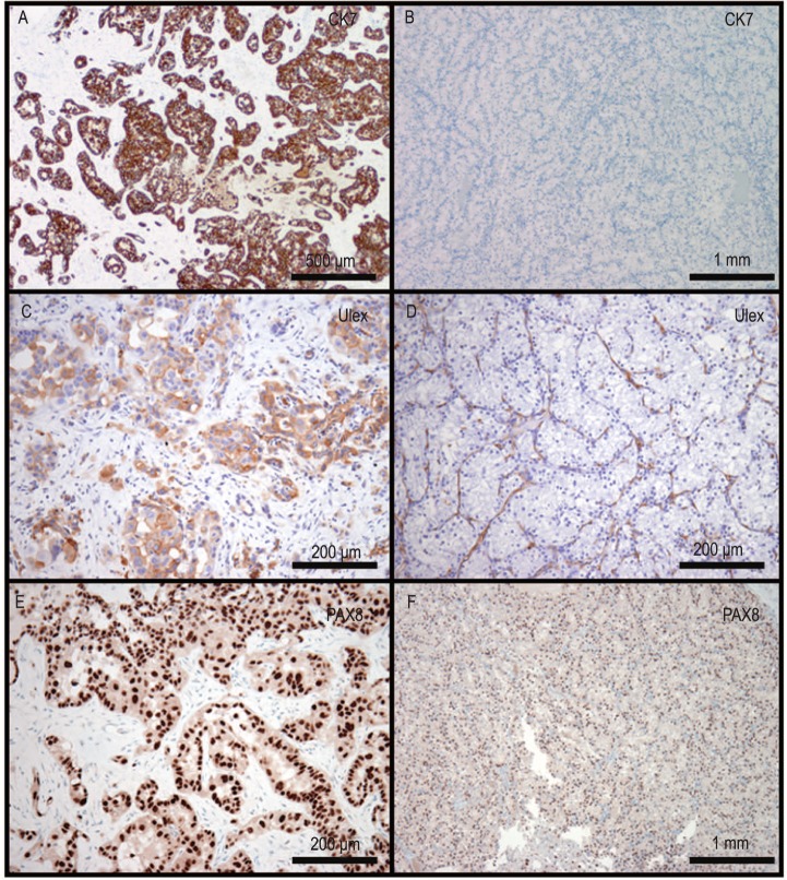
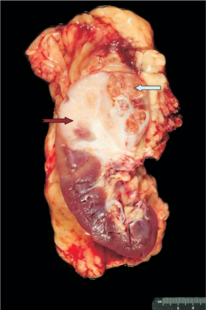
We report the case of a 67-year-old female who presented with a large renal mass. Gross examination of the nephrectomy specimen demonstrated a 6-cm renal mass that invaded into the renal sinus and perinephric fat. Histologic examination revealed two distinct tumor types. The first type was a conventional (clear cell) renal cell carcinoma that was of low nuclear grade and comprised the minority of the overall tumor. The second type was a high-grade collecting duct carcinoma with glandular/tubular differentiation and composed the majority of the tumor. Immunohistochemical studies demonstrated distinctive patterns of the two tumor types, thus confirming two distinct lineages. Five months postoperatively, the patient developed metastasis to the lungs and right hilar lymph node region. A fine needle aspiration of a lung nodule demonstrated a metastatic, poorly differentiated carcinoma, similar to the collecting duct carcinoma component in the kidney. Collision tumors of the kidney are rare with fewer than 10 cases reported in the literature. Our report further expands the spectrum of this rare phenomenon.
期刊介绍:
In July 2008, Landes Bioscience and Sun Yat-sen University Cancer Center began co-publishing the international, English-language version of AI ZHENG or the Chinese Journal of Cancer (CJC). CJC publishes original research, reviews, extra views, perspectives, supplements, and spotlights in all areas of cancer research. The primary criteria for publication in CJC are originality, outstanding scientific merit, and general interest. The Editorial Board is composed of members from around the world, who will strive to maintain the highest standards for excellence in order to generate a valuable resource for an international readership.
 求助内容:
求助内容: 应助结果提醒方式:


