S Weinberger, M Bäder, C Scheurig-Münkler, S Hinz, J Neymeyer, K Miller, C Kempkensteffen
{"title":"Optimizing evaluation of split renal function in a living kidney donor using scintigraphy and calculation of the geometric mean: a case report.","authors":"S Weinberger, M Bäder, C Scheurig-Münkler, S Hinz, J Neymeyer, K Miller, C Kempkensteffen","doi":"10.1159/000358007","DOIUrl":null,"url":null,"abstract":"<p><p>Within the evaluation process of living kidney donors, split renal function is usually evaluated by renal scintigraphy. Since split renal function measured by conventional posterior scans depends on the position of the kidney, actual suitable donors may be rejected because of an inaccurate examination technique. We report the case of a 28-year-old male living kidney donor. Due to a complex vascular anatomy of the right kidney, only his left kidney was considered eligible for transplantation. In conventional posterior Tc99m-mercapto-acetyltriglycine scintigraphy, the left kidney had a relative function of 60%. A second scintigraphy using anterior and posterior dimercaptosuccinic acid scans with calculation of the geometric mean showed an adapted relative function of the left kidney of 53%, now meeting the inclusion criteria for living kidney donation. This case shows that the geometric mean method using simultaneous anterior and posterior views obtained with a dual-head gamma camera can be a very helpful approach to determine split renal function of potential living kidney donors. Further investigation is necessary to prove the benefit of a general bilateral scan before living kidney donation. </p>","PeriodicalId":89663,"journal":{"name":"Case reports in nephrology and urology","volume":"4 1","pages":"1-4"},"PeriodicalIF":0.0000,"publicationDate":"2014-01-10","publicationTypes":"Journal Article","fieldsOfStudy":null,"isOpenAccess":false,"openAccessPdf":"https://sci-hub-pdf.com/10.1159/000358007","citationCount":"6","resultStr":null,"platform":"Semanticscholar","paperid":null,"PeriodicalName":"Case reports in nephrology and urology","FirstCategoryId":"1085","ListUrlMain":"https://doi.org/10.1159/000358007","RegionNum":0,"RegionCategory":null,"ArticlePicture":[],"TitleCN":null,"AbstractTextCN":null,"PMCID":null,"EPubDate":"2014/1/1 0:00:00","PubModel":"eCollection","JCR":"","JCRName":"","Score":null,"Total":0}
引用次数: 6
Abstract
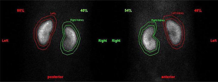
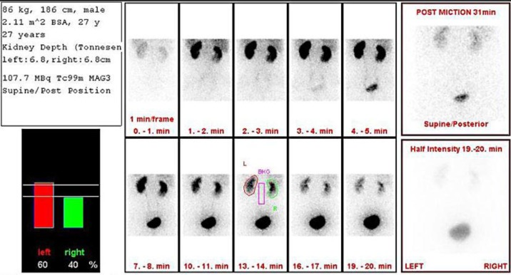
Within the evaluation process of living kidney donors, split renal function is usually evaluated by renal scintigraphy. Since split renal function measured by conventional posterior scans depends on the position of the kidney, actual suitable donors may be rejected because of an inaccurate examination technique. We report the case of a 28-year-old male living kidney donor. Due to a complex vascular anatomy of the right kidney, only his left kidney was considered eligible for transplantation. In conventional posterior Tc99m-mercapto-acetyltriglycine scintigraphy, the left kidney had a relative function of 60%. A second scintigraphy using anterior and posterior dimercaptosuccinic acid scans with calculation of the geometric mean showed an adapted relative function of the left kidney of 53%, now meeting the inclusion criteria for living kidney donation. This case shows that the geometric mean method using simultaneous anterior and posterior views obtained with a dual-head gamma camera can be a very helpful approach to determine split renal function of potential living kidney donors. Further investigation is necessary to prove the benefit of a general bilateral scan before living kidney donation.

 求助内容:
求助内容: 应助结果提醒方式:


