Varun Puvanesarajah, Ioan A Lina, Jason A Liauw, Wesley Hsu, Peter C Burger, Timothy F Witham
{"title":"Desmoid Tumor Formation following Posterior Spinal Instrumentation Placement.","authors":"Varun Puvanesarajah, Ioan A Lina, Jason A Liauw, Wesley Hsu, Peter C Burger, Timothy F Witham","doi":"10.1055/s-0033-1357356","DOIUrl":null,"url":null,"abstract":"<p><p>Study Design Case report. Objective The objective of the article is to illustrate a case of desmoid tumor (DT) formation after posterior instrumentation of the thoracic spine. Methods A 57-year-old woman presented with lower extremity clumsiness, balance, and ambulation difficulty resulting from spinal cord compression due to an upper thoracic atypical vertebral hemangioma. Ten months after undergoing embolization, resection, and placement of instrumentation for this lesion, the patient developed a growing mass at the rostral end of the incision. Biopsy revealed desmoid fibromatosis. The mass was removed via an en bloc resection. Histology revealed an infiltrative DT above the laminectomy site abutting the instrumentation. Results At 2-year follow-up, there was no evidence of recurrence of the tumor. Conclusion Paraspinal DTs have been reported in the literature to develop after surgical procedures of the spine. Often times, patients attribute swelling or fullness at the site of their surgery to scar tissue formation or instrumentation. One must consider the possibility of a DT in the setting of reported surgical site fullness or mass after spine surgery. It is thought that postoperative inflammation present in the surgical bed may promote formation of DTs. Instrumentation may also contribute to inflammation and increase the likelihood of developing a DT. Generous margins must be taken to prevent recurrence. </p>","PeriodicalId":89675,"journal":{"name":"Evidence-based spine-care journal","volume":"4 2","pages":"137-42"},"PeriodicalIF":0.0000,"publicationDate":"2013-10-01","publicationTypes":"Journal Article","fieldsOfStudy":null,"isOpenAccess":false,"openAccessPdf":"https://sci-hub-pdf.com/10.1055/s-0033-1357356","citationCount":"9","resultStr":null,"platform":"Semanticscholar","paperid":null,"PeriodicalName":"Evidence-based spine-care journal","FirstCategoryId":"1085","ListUrlMain":"https://doi.org/10.1055/s-0033-1357356","RegionNum":0,"RegionCategory":null,"ArticlePicture":[],"TitleCN":null,"AbstractTextCN":null,"PMCID":null,"EPubDate":"","PubModel":"","JCR":"","JCRName":"","Score":null,"Total":0}
引用次数: 9
Abstract
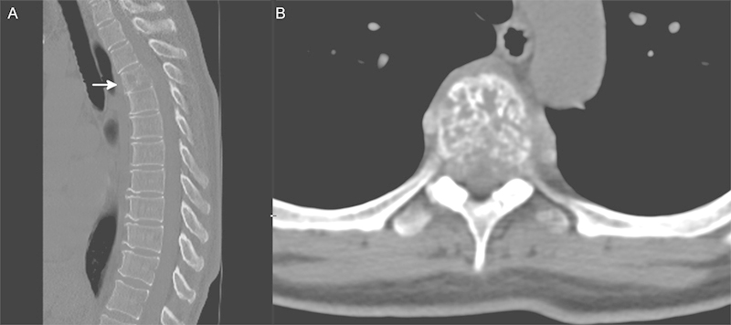
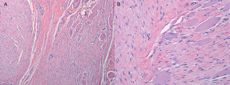
Study Design Case report. Objective The objective of the article is to illustrate a case of desmoid tumor (DT) formation after posterior instrumentation of the thoracic spine. Methods A 57-year-old woman presented with lower extremity clumsiness, balance, and ambulation difficulty resulting from spinal cord compression due to an upper thoracic atypical vertebral hemangioma. Ten months after undergoing embolization, resection, and placement of instrumentation for this lesion, the patient developed a growing mass at the rostral end of the incision. Biopsy revealed desmoid fibromatosis. The mass was removed via an en bloc resection. Histology revealed an infiltrative DT above the laminectomy site abutting the instrumentation. Results At 2-year follow-up, there was no evidence of recurrence of the tumor. Conclusion Paraspinal DTs have been reported in the literature to develop after surgical procedures of the spine. Often times, patients attribute swelling or fullness at the site of their surgery to scar tissue formation or instrumentation. One must consider the possibility of a DT in the setting of reported surgical site fullness or mass after spine surgery. It is thought that postoperative inflammation present in the surgical bed may promote formation of DTs. Instrumentation may also contribute to inflammation and increase the likelihood of developing a DT. Generous margins must be taken to prevent recurrence.


 求助内容:
求助内容: 应助结果提醒方式:


