Ali Humadi, Brian J C Freeman, Rob J Moore, Stuart Callary, Klas Halldin, Vikram David, William Maclaurin, Paul Tauro, Mark Schoenwaelder
{"title":"A comparison of radiostereometric analysis and computed tomography for the assessment of lumbar spinal fusion in a sheep model.","authors":"Ali Humadi, Brian J C Freeman, Rob J Moore, Stuart Callary, Klas Halldin, Vikram David, William Maclaurin, Paul Tauro, Mark Schoenwaelder","doi":"10.1055/s-0033-1357359","DOIUrl":null,"url":null,"abstract":"<p><p>Study Design Prospective animal study. Objective The aim of this animal study is to evaluate the accuracy of radiostereometric analysis (RSA) compared with computed tomographic (CT) scan in the assessment of spinal fusion after anterior lumbar interbody fusion (ALIF) using histology as a gold standard. Methods Three non-adjacent ALIFs (L1-L2, L3-L4, and L5-L6) were performed in nine sheep. The sheep were divided into three groups of three sheep. All the animals were humanely killed immediately after having the last scheduled RSA. The lumbar spine was removed and in vitro fine cut CT and histopathology were performed. Results Using histological assessment as the gold standard for assessing fusion, RSA demonstrated better results (100% sensitivity and 66.7% specificity; positive predictive value [PPV] = 27.3%, negative predictive value [NPV] =100.0%) compared with CT (66.7% sensitivity and 60.0% specificity [PPV = 16.7%, NPV = 93.8%]). Conclusions RSA demonstrated higher sensitivity and specificity when compared with CT. Furthermore, RSA has the advantage of much lower radiation exposure compared with fine cut CT. Further studies are required to see if RSA remains superior to CT scan for the assessment spinal fusion in the clinical setting. [Table: see text]. </p>","PeriodicalId":89675,"journal":{"name":"Evidence-based spine-care journal","volume":"4 2","pages":"78-89"},"PeriodicalIF":0.0000,"publicationDate":"2013-10-01","publicationTypes":"Journal Article","fieldsOfStudy":null,"isOpenAccess":false,"openAccessPdf":"https://sci-hub-pdf.com/10.1055/s-0033-1357359","citationCount":"9","resultStr":null,"platform":"Semanticscholar","paperid":null,"PeriodicalName":"Evidence-based spine-care journal","FirstCategoryId":"1085","ListUrlMain":"https://doi.org/10.1055/s-0033-1357359","RegionNum":0,"RegionCategory":null,"ArticlePicture":[],"TitleCN":null,"AbstractTextCN":null,"PMCID":null,"EPubDate":"","PubModel":"","JCR":"","JCRName":"","Score":null,"Total":0}
引用次数: 9
Abstract
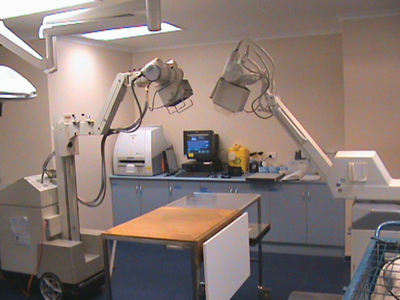
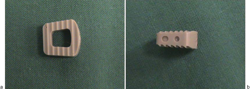
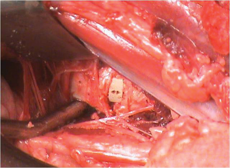
Study Design Prospective animal study. Objective The aim of this animal study is to evaluate the accuracy of radiostereometric analysis (RSA) compared with computed tomographic (CT) scan in the assessment of spinal fusion after anterior lumbar interbody fusion (ALIF) using histology as a gold standard. Methods Three non-adjacent ALIFs (L1-L2, L3-L4, and L5-L6) were performed in nine sheep. The sheep were divided into three groups of three sheep. All the animals were humanely killed immediately after having the last scheduled RSA. The lumbar spine was removed and in vitro fine cut CT and histopathology were performed. Results Using histological assessment as the gold standard for assessing fusion, RSA demonstrated better results (100% sensitivity and 66.7% specificity; positive predictive value [PPV] = 27.3%, negative predictive value [NPV] =100.0%) compared with CT (66.7% sensitivity and 60.0% specificity [PPV = 16.7%, NPV = 93.8%]). Conclusions RSA demonstrated higher sensitivity and specificity when compared with CT. Furthermore, RSA has the advantage of much lower radiation exposure compared with fine cut CT. Further studies are required to see if RSA remains superior to CT scan for the assessment spinal fusion in the clinical setting. [Table: see text].

 求助内容:
求助内容: 应助结果提醒方式:


