{"title":"The Pathophysiology of Primary Hip Osteoarthritis may Originate from Bone Alterations.","authors":"Mikio Kamimura, Yukio Nakamura, Shota Ikegami, Keijiro Mukaiyama, Shigeharu Uchiyama, Hiroyuki Kato","doi":"10.2174/1874312920130930003","DOIUrl":null,"url":null,"abstract":"<p><strong>Objectives: </strong>The aim of this study was to investigate whether bone alterations detected by hip magnetic resonance imaging (MRI) were associated with subsequent primary hip OA.</p><p><strong>Methods: </strong>We enrolled 7 patients with hip joint pain from their first visit, at which hip joints were classified as grade 0 or I on the Kellgren-Lawrence grading scale. Plain radiographs and magnetic resonance imaging (MRI) were performed on all cases, and pain was assessed with the Denis pain scale. Average age, height, weight, body mass index, bone mineral density (L1-4), central edge angle, Sharp's angle, and acetabular hip index were calculated.</p><p><strong>Results: </strong>Within two months of the onset of pain, 4 of the 7 cases showed broad bone signal changes, while 3 cases showed local signal changes in the proximal femur on hip MRI. Three to 6 months after the onset of pain, in all patients whose pain was much improved, plain radiographs showed progression to further-stage OA.</p><p><strong>Conclusion: </strong>Our findings suggest that bone abnormalities in the proximal femur might be involved in the pathogenesis of primary hip OA.</p>","PeriodicalId":39124,"journal":{"name":"Open Rheumatology Journal","volume":"7 ","pages":"112-8"},"PeriodicalIF":0.0000,"publicationDate":"2013-11-29","publicationTypes":"Journal Article","fieldsOfStudy":null,"isOpenAccess":false,"openAccessPdf":"https://ftp.ncbi.nlm.nih.gov/pub/pmc/oa_pdf/a0/91/TORJ-7-112.PMC3866704.pdf","citationCount":"11","resultStr":null,"platform":"Semanticscholar","paperid":null,"PeriodicalName":"Open Rheumatology Journal","FirstCategoryId":"1085","ListUrlMain":"https://doi.org/10.2174/1874312920130930003","RegionNum":0,"RegionCategory":null,"ArticlePicture":[],"TitleCN":null,"AbstractTextCN":null,"PMCID":null,"EPubDate":"2013/1/1 0:00:00","PubModel":"eCollection","JCR":"Q4","JCRName":"Medicine","Score":null,"Total":0}
引用次数: 11
Abstract
Objectives: The aim of this study was to investigate whether bone alterations detected by hip magnetic resonance imaging (MRI) were associated with subsequent primary hip OA.
Methods: We enrolled 7 patients with hip joint pain from their first visit, at which hip joints were classified as grade 0 or I on the Kellgren-Lawrence grading scale. Plain radiographs and magnetic resonance imaging (MRI) were performed on all cases, and pain was assessed with the Denis pain scale. Average age, height, weight, body mass index, bone mineral density (L1-4), central edge angle, Sharp's angle, and acetabular hip index were calculated.
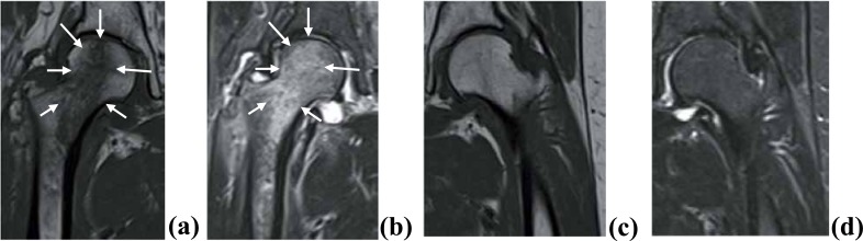
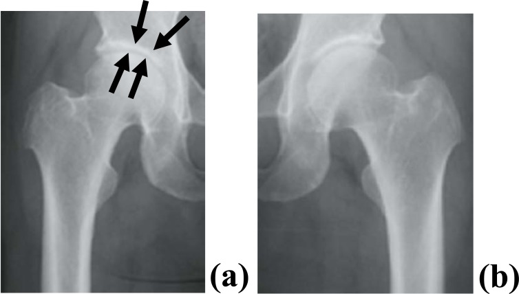
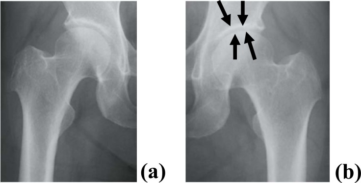
Results: Within two months of the onset of pain, 4 of the 7 cases showed broad bone signal changes, while 3 cases showed local signal changes in the proximal femur on hip MRI. Three to 6 months after the onset of pain, in all patients whose pain was much improved, plain radiographs showed progression to further-stage OA.
Conclusion: Our findings suggest that bone abnormalities in the proximal femur might be involved in the pathogenesis of primary hip OA.
期刊介绍:
ENTHAM Open publishes a number of peer-reviewed, open access journals. These free-to-view online journals cover all major disciplines of science, medicine, technology and social sciences. BENTHAM Open provides researchers a platform to rapidly publish their research in a good-quality peer-reviewed journal. All peer-reviewed accepted submissions meeting high research and ethical standards are published with free access to all.

 求助内容:
求助内容: 应助结果提醒方式:


