Adarsh Shankar, Sanath Kumar, A S M Iskander, Nadimpalli R S Varma, Branislava Janic, Ana deCarvalho, Tom Mikkelsen, Joseph A Frank, Meser M Ali, Robert A Knight, Stephen Brown, Ali S Arbab
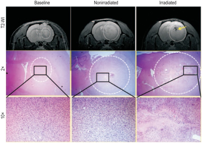
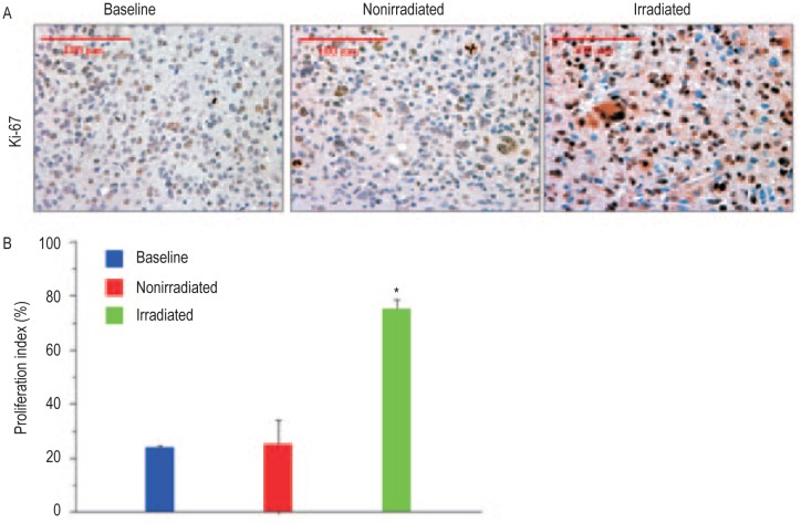
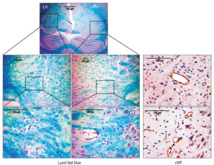
{"title":"Subcurative radiation significantly increases cell proliferation, invasion, and migration of primary glioblastoma multiforme in vivo.","authors":"Adarsh Shankar, Sanath Kumar, A S M Iskander, Nadimpalli R S Varma, Branislava Janic, Ana deCarvalho, Tom Mikkelsen, Joseph A Frank, Meser M Ali, Robert A Knight, Stephen Brown, Ali S Arbab","doi":"10.5732/cjc.013.10095","DOIUrl":null,"url":null,"abstract":"<p><p>Tumor cell proliferation, infiltration, migration, and neovascularization are known causes of treatment resistance in glioblastoma multiforme (GBM). The purpose of this study was to determine the effect of radiation on the growth characteristics of primary human GBM developed in a nude rat. Primary GBM cells grown from explanted GBM tissues were implanted orthotopically in nude rats. Tumor growth was confirmed by magnetic resonance imaging on day 77 (baseline) after implantation. The rats underwent irradiation to a dose of 50 Gy delivered subcuratively on day 84 postimplantation (n = 8), or underwent no radiation (n = 8). Brain tissues were obtained on day 112 (nonirradiated) or day 133 (irradiated). Immunohistochemistry was performed to determine tumor cell proliferation (Ki-67) and to assess the expression of infiltration marker (matrix metalloproteinase-2, MMP-2) and cell migration marker (CD44). Tumor neovascularization was assessed by microvessel density using von-Willebrand factor (vWF) staining. Magnetic resonance imaging showed well-developed, infiltrative tumors in 11 weeks postimplantation. The proportion of Ki-67-positive cells in tumors undergoing radiation was (71 +/- 15)% compared with (25 +/- 12)% in the nonirradiated group (P = 0.02). The number of MMP-2-positive areas and proportion of CD44-positive cells were also high in tumors receiving radiation, indicating great invasion and infiltration. Microvessel density analysis did not show a significant difference between nonirradiated and irradiated tumors. Taken together, we found that subcurative radiation significantly increased proliferation, invasion, and migration of primary GBM. Our study provides insights into possible mechanisms of treatment resistance following radiation therapy for GBM. </p>","PeriodicalId":10034,"journal":{"name":"癌症","volume":"33 3","pages":"148-58"},"PeriodicalIF":0.0000,"publicationDate":"2014-03-01","publicationTypes":"Journal Article","fieldsOfStudy":null,"isOpenAccess":false,"openAccessPdf":"https://ftp.ncbi.nlm.nih.gov/pub/pmc/oa_pdf/2d/7e/cjc-33-03-148.PMC3966215.pdf","citationCount":"33","resultStr":null,"platform":"Semanticscholar","paperid":null,"PeriodicalName":"癌症","FirstCategoryId":"3","ListUrlMain":"https://doi.org/10.5732/cjc.013.10095","RegionNum":0,"RegionCategory":null,"ArticlePicture":[],"TitleCN":null,"AbstractTextCN":null,"PMCID":null,"EPubDate":"2013/9/9 0:00:00","PubModel":"Epub","JCR":"Q","JCRName":"Medicine","Score":null,"Total":0}
引用次数: 33
Abstract
Tumor cell proliferation, infiltration, migration, and neovascularization are known causes of treatment resistance in glioblastoma multiforme (GBM). The purpose of this study was to determine the effect of radiation on the growth characteristics of primary human GBM developed in a nude rat. Primary GBM cells grown from explanted GBM tissues were implanted orthotopically in nude rats. Tumor growth was confirmed by magnetic resonance imaging on day 77 (baseline) after implantation. The rats underwent irradiation to a dose of 50 Gy delivered subcuratively on day 84 postimplantation (n = 8), or underwent no radiation (n = 8). Brain tissues were obtained on day 112 (nonirradiated) or day 133 (irradiated). Immunohistochemistry was performed to determine tumor cell proliferation (Ki-67) and to assess the expression of infiltration marker (matrix metalloproteinase-2, MMP-2) and cell migration marker (CD44). Tumor neovascularization was assessed by microvessel density using von-Willebrand factor (vWF) staining. Magnetic resonance imaging showed well-developed, infiltrative tumors in 11 weeks postimplantation. The proportion of Ki-67-positive cells in tumors undergoing radiation was (71 +/- 15)% compared with (25 +/- 12)% in the nonirradiated group (P = 0.02). The number of MMP-2-positive areas and proportion of CD44-positive cells were also high in tumors receiving radiation, indicating great invasion and infiltration. Microvessel density analysis did not show a significant difference between nonirradiated and irradiated tumors. Taken together, we found that subcurative radiation significantly increased proliferation, invasion, and migration of primary GBM. Our study provides insights into possible mechanisms of treatment resistance following radiation therapy for GBM.
期刊介绍:
In July 2008, Landes Bioscience and Sun Yat-sen University Cancer Center began co-publishing the international, English-language version of AI ZHENG or the Chinese Journal of Cancer (CJC). CJC publishes original research, reviews, extra views, perspectives, supplements, and spotlights in all areas of cancer research. The primary criteria for publication in CJC are originality, outstanding scientific merit, and general interest. The Editorial Board is composed of members from around the world, who will strive to maintain the highest standards for excellence in order to generate a valuable resource for an international readership.

 求助内容:
求助内容: 应助结果提醒方式:


