Akram M Asbeutah, Yousif Y Bakir, Nayanatara Swamy, Abdul Aziz A Absuetah, Muna A Abu-Asi, Prem Sharma
{"title":"Subject body mass index affects Doppler waveform in celiac artery by duplex ultrasound.","authors":"Akram M Asbeutah, Yousif Y Bakir, Nayanatara Swamy, Abdul Aziz A Absuetah, Muna A Abu-Asi, Prem Sharma","doi":"10.2174/1874192401307010040","DOIUrl":null,"url":null,"abstract":"<p><strong>Objective: </strong>The aim of this study is to evaluate the effect of body mass index (BMI) on peak systolic velocity (PSV) recording in the celiac artery (CA).</p><p><strong>Subjects & methods: </strong>Forty male participants were entered prospectively into the study. The subjects were divided into two groups according to their body mass index. Group A included subjects with BMI ≤25 Kg/m(2) and those in group B with BMI >25 Kg/m(2). The diameter and PSV at the origin of CA of subjects in both groups were recorded while the subject positioned in supine and during expiration phase and fasted for 4 hours using duplex ultrasound. Both groups were matched for age and sex. Independent Student's t-test was used to test if there is any statistical significance between diameter and PSV in both groups.</p><p><strong>Results: </strong>Group A's, average age (year, ±SD) was 29.35±1.35 and average BMI (Kg/m(2), ±SD) was 23.1±1.60. Group B's, average age was 30±2.1 and their average BMI was 31±5.1. The average diameter (cm, ±SD) of CA in group A was 0.66±0.076 and in group B was 0.80±0.066. However, the average PSV (cm/s, ±SD) was 117±28.1 in group A and 102±12.4 in group B. Independent student t-test showed statistical significance between both groups for the diameter (p=0.005) and just reached statistical significance for PSV (p=0.049).</p><p><strong>Conclusion: </strong>Subjects with higher BMI showed reduced PSV due to a larger CA diameter and probably due to more fatty tissue accumulation around the CA origin.</p>","PeriodicalId":504447,"journal":{"name":"The Open Cardiovascular Medicine Journal","volume":"7 ","pages":"40-5"},"PeriodicalIF":0.0000,"publicationDate":"2013-04-30","publicationTypes":"Journal Article","fieldsOfStudy":null,"isOpenAccess":false,"openAccessPdf":"https://ftp.ncbi.nlm.nih.gov/pub/pmc/oa_pdf/34/b8/TOCMJ-7-40.PMC3681032.pdf","citationCount":"5","resultStr":null,"platform":"Semanticscholar","paperid":null,"PeriodicalName":"The Open Cardiovascular Medicine Journal","FirstCategoryId":"1085","ListUrlMain":"https://doi.org/10.2174/1874192401307010040","RegionNum":0,"RegionCategory":null,"ArticlePicture":[],"TitleCN":null,"AbstractTextCN":null,"PMCID":null,"EPubDate":"2013/1/1 0:00:00","PubModel":"Print","JCR":"","JCRName":"","Score":null,"Total":0}
引用次数: 5
Abstract
Objective: The aim of this study is to evaluate the effect of body mass index (BMI) on peak systolic velocity (PSV) recording in the celiac artery (CA).
Subjects & methods: Forty male participants were entered prospectively into the study. The subjects were divided into two groups according to their body mass index. Group A included subjects with BMI ≤25 Kg/m(2) and those in group B with BMI >25 Kg/m(2). The diameter and PSV at the origin of CA of subjects in both groups were recorded while the subject positioned in supine and during expiration phase and fasted for 4 hours using duplex ultrasound. Both groups were matched for age and sex. Independent Student's t-test was used to test if there is any statistical significance between diameter and PSV in both groups.
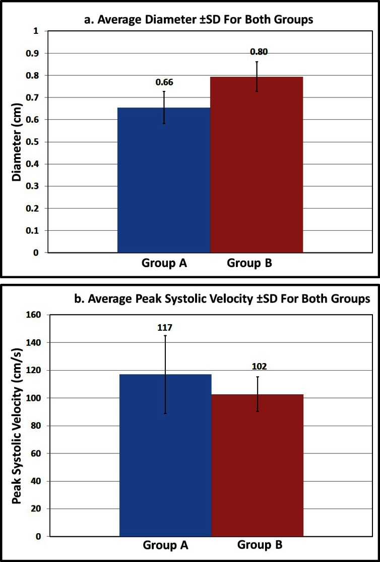
Results: Group A's, average age (year, ±SD) was 29.35±1.35 and average BMI (Kg/m(2), ±SD) was 23.1±1.60. Group B's, average age was 30±2.1 and their average BMI was 31±5.1. The average diameter (cm, ±SD) of CA in group A was 0.66±0.076 and in group B was 0.80±0.066. However, the average PSV (cm/s, ±SD) was 117±28.1 in group A and 102±12.4 in group B. Independent student t-test showed statistical significance between both groups for the diameter (p=0.005) and just reached statistical significance for PSV (p=0.049).
Conclusion: Subjects with higher BMI showed reduced PSV due to a larger CA diameter and probably due to more fatty tissue accumulation around the CA origin.

 求助内容:
求助内容: 应助结果提醒方式:


