{"title":"Ultrasonographic imaging for structural characterization of renal affections and diagnosis of associated chronic renal failure in 10 dogs.","authors":"Vijay Kumar, Adarsh Kumar, A C Varshney","doi":"10.5402/2011/901713","DOIUrl":null,"url":null,"abstract":"<p><p>The present study comprises of 10 dogs of either sex with primary indication of azotaemia. All the dogs were subjected to detailed clinical, haematobiochemical, urinalysis, and microbiological examination along with radiographical and ultrasonographical examination. Based on the ultrasonographic structural abnormalities, the different renal affections associated with CRF in majority of dogs were diagnosed. The different affections included \"end-stage\" kidneys (n = 4), hydronephrosis (n = 1), renomegaly (n = 1), nephritis (n = 1), nephrolithiasis (n = 1), nephrocalcinosis (n = 1), and renal cyst (n = 1). The significant ultrasonographic features in these affections included small kidneys with loss of corticomedullary demarcation (\"end-stage\" kidneys); increased cortical echogenicity (nephritis); dilation of the renal pelvis, separation of the central renal sinus with anechoic space, atrophy of renal medulla, (hydronephrosis); enlarged kidneys with increased overall echogenicity of renal cortex (renomegaly and associated nephritis); hyperechoic-mineralized structure with shadowing (nephrolithiasis); diffuse, small, multiple hyperechoic structures in the renal parenchyma with distal acoustic shadowing (nephrocalcinosis); small spherical intercortical anechoic structures fluid (renal cysts). In the present study, ultrasound proved to be a quick, convenient, and sensitive modality in detecting alterations in renal size and parenchymal architecture. All the dogs so diagnosed with CRF were rendered conservative medical treatment to control clinical signs of uraemia; maintain adequate fluid, electrolyte, and acid/base balance; provide adequate nutrition; minimize progression of renal failure.</p>","PeriodicalId":89682,"journal":{"name":"ISRN veterinary science","volume":"2011 ","pages":"901713"},"PeriodicalIF":0.0000,"publicationDate":"2011-12-25","publicationTypes":"Journal Article","fieldsOfStudy":null,"isOpenAccess":false,"openAccessPdf":"https://sci-hub-pdf.com/10.5402/2011/901713","citationCount":"8","resultStr":null,"platform":"Semanticscholar","paperid":null,"PeriodicalName":"ISRN veterinary science","FirstCategoryId":"1085","ListUrlMain":"https://doi.org/10.5402/2011/901713","RegionNum":0,"RegionCategory":null,"ArticlePicture":[],"TitleCN":null,"AbstractTextCN":null,"PMCID":null,"EPubDate":"2011/1/1 0:00:00","PubModel":"Print","JCR":"","JCRName":"","Score":null,"Total":0}
引用次数: 8
Abstract
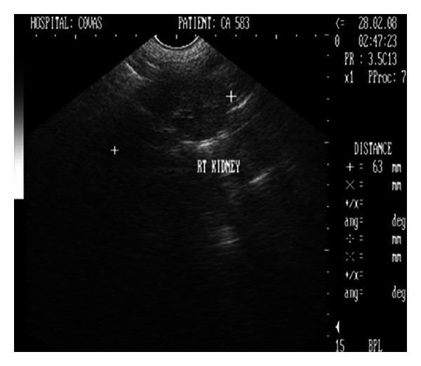
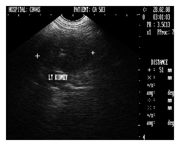
The present study comprises of 10 dogs of either sex with primary indication of azotaemia. All the dogs were subjected to detailed clinical, haematobiochemical, urinalysis, and microbiological examination along with radiographical and ultrasonographical examination. Based on the ultrasonographic structural abnormalities, the different renal affections associated with CRF in majority of dogs were diagnosed. The different affections included "end-stage" kidneys (n = 4), hydronephrosis (n = 1), renomegaly (n = 1), nephritis (n = 1), nephrolithiasis (n = 1), nephrocalcinosis (n = 1), and renal cyst (n = 1). The significant ultrasonographic features in these affections included small kidneys with loss of corticomedullary demarcation ("end-stage" kidneys); increased cortical echogenicity (nephritis); dilation of the renal pelvis, separation of the central renal sinus with anechoic space, atrophy of renal medulla, (hydronephrosis); enlarged kidneys with increased overall echogenicity of renal cortex (renomegaly and associated nephritis); hyperechoic-mineralized structure with shadowing (nephrolithiasis); diffuse, small, multiple hyperechoic structures in the renal parenchyma with distal acoustic shadowing (nephrocalcinosis); small spherical intercortical anechoic structures fluid (renal cysts). In the present study, ultrasound proved to be a quick, convenient, and sensitive modality in detecting alterations in renal size and parenchymal architecture. All the dogs so diagnosed with CRF were rendered conservative medical treatment to control clinical signs of uraemia; maintain adequate fluid, electrolyte, and acid/base balance; provide adequate nutrition; minimize progression of renal failure.

 求助内容:
求助内容: 应助结果提醒方式:


