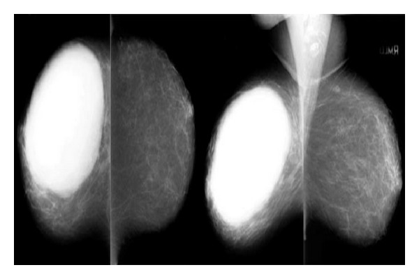
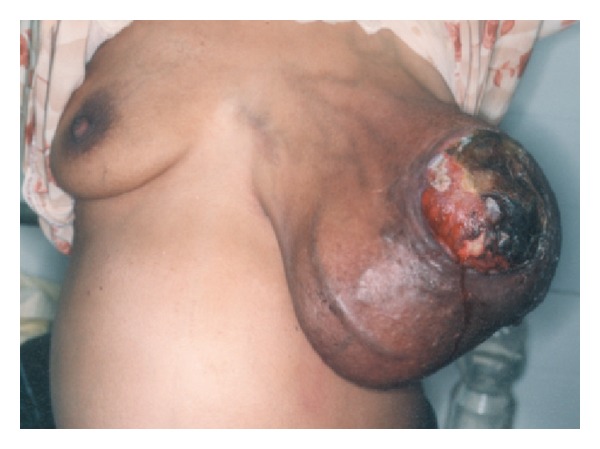
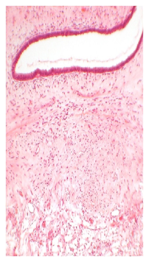
{"title":"Phyllodes tumor of breast: a review article.","authors":"Shashi Prakash Mishra, Satyendra Kumar Tiwary, Manjaree Mishra, Ajay Kumar Khanna","doi":"10.1155/2013/361469","DOIUrl":null,"url":null,"abstract":"<p><p>Introduction. Phyllodes tumours are rare fibroepithelial lesions. Accurate preoperative pathological diagnosis allows correct surgical planning and avoidance of reoperation. Treatment can be either wide local excision or mastectomy to achieve histologically clear margins. Discussion. The exact aetiology of phyllodes tumour and its relationship with fibroadenoma are unclear. Women aged between 35 and 55 years are commonly involved. The median tumour size is 4 cm but can grow even larger having dilated veins and a blue discoloration over skin. Palpable axillary lymphadenopathy can be identified in up to 10-15% of patients but <1% had pathological positive nodes. Mammography and ultrasonography are main imaging modalities. Cytologically the presence of both epithelial and stromal elements supports the diagnosis. The value of FNAC in diagnosis of phyllodes tumour remains controversial, but core needle biopsy has high sensitivity and negative predictive value. Surgical management is the mainstay and local recurrence in phyllodes tumours has been associated with inadequate local excision. The role of adjuvant radiotherapy and chemotherapy remains uncertain and use of hormonal therapy has not been fully investigated. Conclusion. The preoperative diagnosis and proper management are crucial in phyllodes tumours because of their tendency to recur and malignant potential in some of these tumours.</p>","PeriodicalId":89400,"journal":{"name":"ISRN surgery","volume":"2013 ","pages":"361469"},"PeriodicalIF":0.0000,"publicationDate":"2013-01-01","publicationTypes":"Journal Article","fieldsOfStudy":null,"isOpenAccess":false,"openAccessPdf":"https://www.ncbi.nlm.nih.gov/pmc/articles/PMC3615633/pdf/","citationCount":"0","resultStr":null,"platform":"Semanticscholar","paperid":null,"PeriodicalName":"ISRN surgery","FirstCategoryId":"1085","ListUrlMain":"https://doi.org/10.1155/2013/361469","RegionNum":0,"RegionCategory":null,"ArticlePicture":[],"TitleCN":null,"AbstractTextCN":null,"PMCID":null,"EPubDate":"2013/3/20 0:00:00","PubModel":"Epub","JCR":"","JCRName":"","Score":null,"Total":0}
引用次数: 0
Abstract
Introduction. Phyllodes tumours are rare fibroepithelial lesions. Accurate preoperative pathological diagnosis allows correct surgical planning and avoidance of reoperation. Treatment can be either wide local excision or mastectomy to achieve histologically clear margins. Discussion. The exact aetiology of phyllodes tumour and its relationship with fibroadenoma are unclear. Women aged between 35 and 55 years are commonly involved. The median tumour size is 4 cm but can grow even larger having dilated veins and a blue discoloration over skin. Palpable axillary lymphadenopathy can be identified in up to 10-15% of patients but <1% had pathological positive nodes. Mammography and ultrasonography are main imaging modalities. Cytologically the presence of both epithelial and stromal elements supports the diagnosis. The value of FNAC in diagnosis of phyllodes tumour remains controversial, but core needle biopsy has high sensitivity and negative predictive value. Surgical management is the mainstay and local recurrence in phyllodes tumours has been associated with inadequate local excision. The role of adjuvant radiotherapy and chemotherapy remains uncertain and use of hormonal therapy has not been fully investigated. Conclusion. The preoperative diagnosis and proper management are crucial in phyllodes tumours because of their tendency to recur and malignant potential in some of these tumours.

 求助内容:
求助内容: 应助结果提醒方式:


