A Bogaert-Buchmann, M Poittevin, C Po, D Dupont, C Sebrié, Y Tomita, A Trandinh, J Seylaz, E Pinard, P Méric, N Kubis, B Gillet
{"title":"Spatial and temporal MRI profile of ischemic tissue after the acute stages of a permanent mouse model of stroke.","authors":"A Bogaert-Buchmann, M Poittevin, C Po, D Dupont, C Sebrié, Y Tomita, A Trandinh, J Seylaz, E Pinard, P Méric, N Kubis, B Gillet","doi":"10.2174/1874440001307010004","DOIUrl":null,"url":null,"abstract":"<p><strong>Object: </strong>To characterize the progression of injured tissue resulting from a permanent focal cerebral ischemia after the acute phase, Magnetic Resonance Imaging (MRI) monitoring was performed on adult male C57BL/6J mice in the subacute stages, and correlated to histological analyses.</p><p><strong>Material and methods: </strong>Lesions were induced by electrocoagulation of the middle cerebral artery. Serial MRI measurements and weighted-images (T2, T1, T2* and Diffusion Tensor Imaging) were performed on a 9.4T scanner. Histological data (Cresyl-Violet staining and laminin-, Iba1- and GFAP-immunostainings) were obtained 1 and 2 weeks after the stroke.</p><p><strong>Results: </strong>Two days after stroke, tissues assumed to correspond to the infarct core, were detected as a hyperintensity signal area in T2-weighted images. One week later, low-intensity signal areas appeared. Longitudinal MRI study showed that these areas remained present over the following week, and was mainly linked to a drop of the T2 relaxation time value in the corresponding tissues. Correlation with histological data and immuno-histochemistry showed that these areas corresponded to microglial cells.</p><p><strong>Conclusion: </strong>The present data provide, for the first time detailed MRI parameters of microglial cells dynamics, allowing its non-invasive monitoring during the chronic stages of a stroke. This could be particularly interesting in regards to emerging anti-inflammatory stroke therapies.</p>","PeriodicalId":37431,"journal":{"name":"Open Neuroimaging Journal","volume":"7 ","pages":"4-14"},"PeriodicalIF":0.0000,"publicationDate":"2013-01-01","publicationTypes":"Journal Article","fieldsOfStudy":null,"isOpenAccess":false,"openAccessPdf":"https://ftp.ncbi.nlm.nih.gov/pub/pmc/oa_pdf/0f/48/TONIJ-7-4.PMC3580904.pdf","citationCount":"6","resultStr":null,"platform":"Semanticscholar","paperid":null,"PeriodicalName":"Open Neuroimaging Journal","FirstCategoryId":"1085","ListUrlMain":"https://doi.org/10.2174/1874440001307010004","RegionNum":0,"RegionCategory":null,"ArticlePicture":[],"TitleCN":null,"AbstractTextCN":null,"PMCID":null,"EPubDate":"2013/2/1 0:00:00","PubModel":"Epub","JCR":"Q4","JCRName":"Medicine","Score":null,"Total":0}
引用次数: 6
Abstract
Object: To characterize the progression of injured tissue resulting from a permanent focal cerebral ischemia after the acute phase, Magnetic Resonance Imaging (MRI) monitoring was performed on adult male C57BL/6J mice in the subacute stages, and correlated to histological analyses.
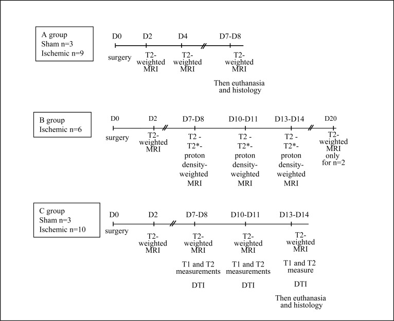
Material and methods: Lesions were induced by electrocoagulation of the middle cerebral artery. Serial MRI measurements and weighted-images (T2, T1, T2* and Diffusion Tensor Imaging) were performed on a 9.4T scanner. Histological data (Cresyl-Violet staining and laminin-, Iba1- and GFAP-immunostainings) were obtained 1 and 2 weeks after the stroke.
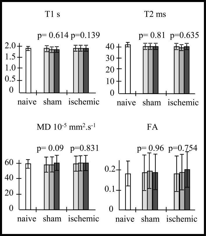
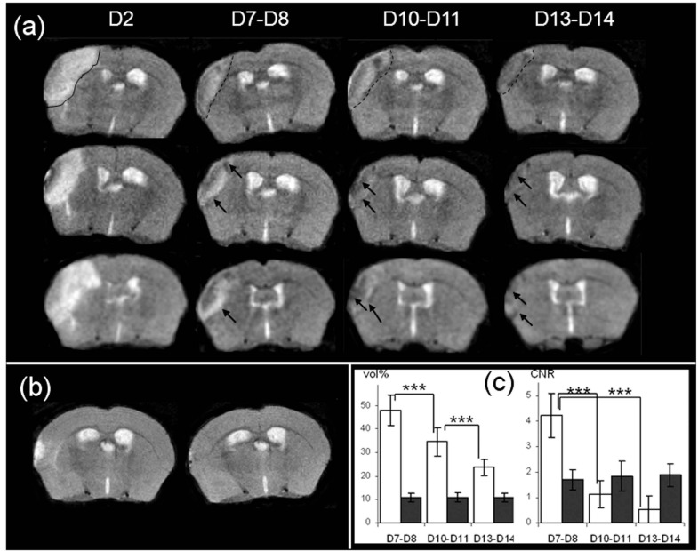
Results: Two days after stroke, tissues assumed to correspond to the infarct core, were detected as a hyperintensity signal area in T2-weighted images. One week later, low-intensity signal areas appeared. Longitudinal MRI study showed that these areas remained present over the following week, and was mainly linked to a drop of the T2 relaxation time value in the corresponding tissues. Correlation with histological data and immuno-histochemistry showed that these areas corresponded to microglial cells.
Conclusion: The present data provide, for the first time detailed MRI parameters of microglial cells dynamics, allowing its non-invasive monitoring during the chronic stages of a stroke. This could be particularly interesting in regards to emerging anti-inflammatory stroke therapies.
期刊介绍:
The Open Neuroimaging Journal is an Open Access online journal, which publishes research articles, reviews/mini-reviews, and letters in all important areas of brain function, structure and organization including neuroimaging, neuroradiology, analysis methods, functional MRI acquisition and physics, brain mapping, macroscopic level of brain organization, computational modeling and analysis, structure-function and brain-behavior relationships, anatomy and physiology, psychiatric diseases and disorders of the nervous system, use of imaging to the understanding of brain pathology and brain abnormalities, cognition and aging, social neuroscience, sensorimotor processing, communication and learning.

 求助内容:
求助内容: 应助结果提醒方式:


