André R Greenidge, Kiana R Hall, Ian R Hambleton, Richelle Thomas, Dougald M Monroe, R Clive Landis
{"title":"Plasmin Activation of Glial Cells through Protease-Activated Receptor 1.","authors":"André R Greenidge, Kiana R Hall, Ian R Hambleton, Richelle Thomas, Dougald M Monroe, R Clive Landis","doi":"10.1155/2013/314709","DOIUrl":null,"url":null,"abstract":"<p><p>The objective of this study was to determine whether plasmin could induce morphological changes in human glial cells via PAR1. Human glioblastoma A172 cells were cultured in the presence of plasmin or the PAR1 specific activating hexapeptide, SFLLRN. Cells were monitored by flow cytometry to detect proteolytic activation of PAR1 receptor. Morphological changes were recorded by photomicroscopy and apoptosis was measured by annexinV staining. Plasmin cleaved the PAR1 receptor on glial cells at 5 minutes (P = 0.02). After 30 minutes, cellular processes had begun to retract from the basal substratum and by 4 hours glial cells had become detached. Similar results were obtained by generating plasmin de novo from plasminogen. Morphological transformation was blocked by plasmin inhibitors aprotinin or epsilon-aminocaproic acid (P = 0.03). Cell viability was unimpaired during early morphological changes, but by 24 hours following plasmin treatment 22% of glial cells were apoptotic. PAR1 activating peptide SFLLRN (but not inactive isomer FSLLRN) promoted analogous glial cell detachment (P = 0.03), proving the role for PAR1 in this process. This study has identified a plasmin/PAR1 axis of glial cell activation, linked to changes in glial cell morophology. This adds to our understanding of pathophysiological disease mechanisms of plasmin and the plasminogen system in neuroinjury.</p>","PeriodicalId":89212,"journal":{"name":"Pathology research international","volume":"2013 ","pages":"314709"},"PeriodicalIF":0.0000,"publicationDate":"2013-01-01","publicationTypes":"Journal Article","fieldsOfStudy":null,"isOpenAccess":false,"openAccessPdf":"https://sci-hub-pdf.com/10.1155/2013/314709","citationCount":"8","resultStr":null,"platform":"Semanticscholar","paperid":null,"PeriodicalName":"Pathology research international","FirstCategoryId":"1085","ListUrlMain":"https://doi.org/10.1155/2013/314709","RegionNum":0,"RegionCategory":null,"ArticlePicture":[],"TitleCN":null,"AbstractTextCN":null,"PMCID":null,"EPubDate":"2013/1/28 0:00:00","PubModel":"Epub","JCR":"","JCRName":"","Score":null,"Total":0}
引用次数: 8
Abstract
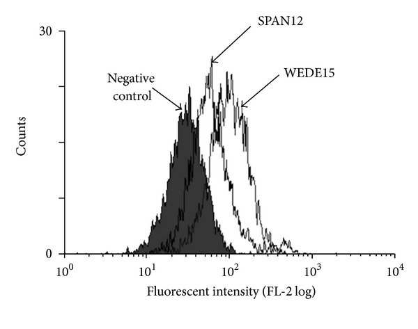
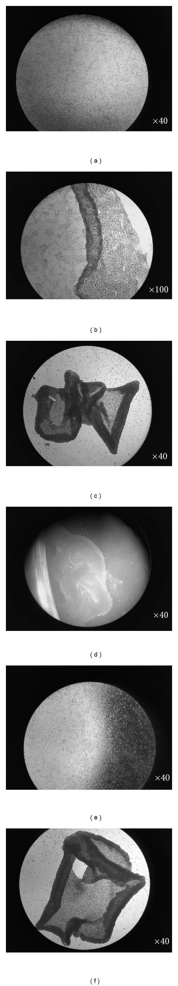
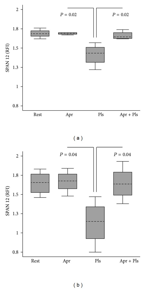
The objective of this study was to determine whether plasmin could induce morphological changes in human glial cells via PAR1. Human glioblastoma A172 cells were cultured in the presence of plasmin or the PAR1 specific activating hexapeptide, SFLLRN. Cells were monitored by flow cytometry to detect proteolytic activation of PAR1 receptor. Morphological changes were recorded by photomicroscopy and apoptosis was measured by annexinV staining. Plasmin cleaved the PAR1 receptor on glial cells at 5 minutes (P = 0.02). After 30 minutes, cellular processes had begun to retract from the basal substratum and by 4 hours glial cells had become detached. Similar results were obtained by generating plasmin de novo from plasminogen. Morphological transformation was blocked by plasmin inhibitors aprotinin or epsilon-aminocaproic acid (P = 0.03). Cell viability was unimpaired during early morphological changes, but by 24 hours following plasmin treatment 22% of glial cells were apoptotic. PAR1 activating peptide SFLLRN (but not inactive isomer FSLLRN) promoted analogous glial cell detachment (P = 0.03), proving the role for PAR1 in this process. This study has identified a plasmin/PAR1 axis of glial cell activation, linked to changes in glial cell morophology. This adds to our understanding of pathophysiological disease mechanisms of plasmin and the plasminogen system in neuroinjury.

 求助内容:
求助内容: 应助结果提醒方式:


