Moraima Morales-Cruz, Giselle M. Flores-Fernández, Myreisa Morales-Cruz, Elsie A. Orellano, José A. Rodriguez-Martinez, Mercedes Ruiz, Kai Griebenow
{"title":"Two-step nanoprecipitation for the production of protein-loaded PLGA nanospheres","authors":"Moraima Morales-Cruz, Giselle M. Flores-Fernández, Myreisa Morales-Cruz, Elsie A. Orellano, José A. Rodriguez-Martinez, Mercedes Ruiz, Kai Griebenow","doi":"10.1016/j.rinphs.2012.11.001","DOIUrl":null,"url":null,"abstract":"<div><p>One of the first methods to encapsulate drugs within polymer nanospheres was developed by Fessi and coworkers in 1989 and consisted of one-step nanoprecipitation based on solvent displacement. However, proteins are poorly encapsulated within polymer nanoparticles using this method because of their limited solubility in organic solvents. To overcome this limitation, we developed a two-step nanoprecipitation method and encapsulated various proteins with high efficiency into poly(lactic-<em>co</em>-glycolic)acid (PLGA) nanospheres (NP). In this method, a protein nanoprecipitation step is used first followed by a second polymer nanoprecipitation step. Two model enzymes, lysozyme and α-chymotrypsin, were used for the optimization of the method. We obtained encapsulation efficiencies of >70%, an amount of buffer-insoluble protein aggregates of typically <2%, and a high residual activity of typically >90%. The optimum conditions identified for lysozyme were used to successfully encapsulate cytochrome <em>c</em>(Cyt-c), an apoptosis-initiating basic protein of similar size, to verify reproducibility of the encapsulation procedure. The size of the Cyt-c loaded-PLGA nanospheres was around 300–400<!--> <!-->nm indicating the potential of the delivery system to passively target tumors. Cell viability studies, using a human cervical cancer cell line (HeLa), demonstrate excellent biocompatibility of the PLGA nanoparticles. PLGA nanoparticles carrying encapsulated Cyt-c were not efficient in causing apoptosis presumably because PLGA nanoparticles are not efficiently taken up by the cells. Future systems will have to be optimized to ascertain efficient cellular uptake of the nanoparticles by, e.g., surface modification with receptor ligands.</p></div>","PeriodicalId":89718,"journal":{"name":"Results in pharma sciences","volume":"2 ","pages":"Pages 79-85"},"PeriodicalIF":0.0000,"publicationDate":"2012-01-01","publicationTypes":"Journal Article","fieldsOfStudy":null,"isOpenAccess":false,"openAccessPdf":"https://sci-hub-pdf.com/10.1016/j.rinphs.2012.11.001","citationCount":"77","resultStr":null,"platform":"Semanticscholar","paperid":null,"PeriodicalName":"Results in pharma sciences","FirstCategoryId":"1085","ListUrlMain":"https://www.sciencedirect.com/science/article/pii/S2211286312000115","RegionNum":0,"RegionCategory":null,"ArticlePicture":[],"TitleCN":null,"AbstractTextCN":null,"PMCID":null,"EPubDate":"","PubModel":"","JCR":"","JCRName":"","Score":null,"Total":0}
引用次数: 77
Abstract
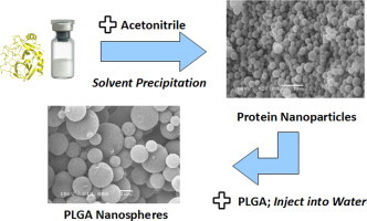
One of the first methods to encapsulate drugs within polymer nanospheres was developed by Fessi and coworkers in 1989 and consisted of one-step nanoprecipitation based on solvent displacement. However, proteins are poorly encapsulated within polymer nanoparticles using this method because of their limited solubility in organic solvents. To overcome this limitation, we developed a two-step nanoprecipitation method and encapsulated various proteins with high efficiency into poly(lactic-co-glycolic)acid (PLGA) nanospheres (NP). In this method, a protein nanoprecipitation step is used first followed by a second polymer nanoprecipitation step. Two model enzymes, lysozyme and α-chymotrypsin, were used for the optimization of the method. We obtained encapsulation efficiencies of >70%, an amount of buffer-insoluble protein aggregates of typically <2%, and a high residual activity of typically >90%. The optimum conditions identified for lysozyme were used to successfully encapsulate cytochrome c(Cyt-c), an apoptosis-initiating basic protein of similar size, to verify reproducibility of the encapsulation procedure. The size of the Cyt-c loaded-PLGA nanospheres was around 300–400 nm indicating the potential of the delivery system to passively target tumors. Cell viability studies, using a human cervical cancer cell line (HeLa), demonstrate excellent biocompatibility of the PLGA nanoparticles. PLGA nanoparticles carrying encapsulated Cyt-c were not efficient in causing apoptosis presumably because PLGA nanoparticles are not efficiently taken up by the cells. Future systems will have to be optimized to ascertain efficient cellular uptake of the nanoparticles by, e.g., surface modification with receptor ligands.


 求助内容:
求助内容: 应助结果提醒方式:


