Marta Lydka, Barbara Bilinska, C Yan Cheng, Dolores D Mruk
{"title":"Tumor necrosis factor α-mediated restructuring of the Sertoli cell barrier in vitro involves matrix metalloprotease 9 (MMP9), membrane-bound intercellular adhesion molecule-1 (ICAM-1) and the actin cytoskeleton.","authors":"Marta Lydka, Barbara Bilinska, C Yan Cheng, Dolores D Mruk","doi":"10.4161/spmg.22602","DOIUrl":null,"url":null,"abstract":"<p><p>The mammalian blood-testis barrier (BTB) restructures throughout spermatogenesis, thereby allowing developing germ cells to enter the adluminal compartment of the seminiferous epithelium. Previous studies have shown pro-inflammatory cytokines such as tumor necrosis factor α (TNFα) and interleukin-1α to be important regulators of Sertoli cell barrier/BTB function in vitro and in vivo. In this study, the effects of TNFα on Sertoli cell barrier function were assessed, with emphasis on changes in proteases and cell adhesion molecules following treatment. By immunoblotting and immunohistochemistry, MMP9 was found to be present in germ cells, localizing by and large to spermatocytes and spermatids in the adult rat testis. Following treatment of Sertoli cells with physiologically relevant consecutive doses of recombinant human TNFα (25 ng/ml), the steady-state levels of active-matrix metalloprotease 9 (MMP9), membrane-bound intercellular adhesion molecule (mICAM-1) and androgen receptor increased significantly. TNFα also downregulated the steady-state level of occludin, in agreement with earlier results that showed TNFα to disrupt Sertoli cell barrier/BTB function. In addition, TNFα affected the filamentous actin cytoskeleton in Sertoli cells, which appeared to be mediated by cortactin, a regulator of actin dynamics. Taken collectively, these findings imply that germ cells may be involved in BTB restructuring via the localized production of TNFα. These results also illustrate that barrier restructuring correlated with an increase in Sertoli cell mICAM-1, suggesting that it may be critical for adhesion as germ cells traverse the \"opened\" BTB.</p>","PeriodicalId":22074,"journal":{"name":"Spermatogenesis","volume":"2 4","pages":"294-303"},"PeriodicalIF":0.0000,"publicationDate":"2012-10-01","publicationTypes":"Journal Article","fieldsOfStudy":null,"isOpenAccess":false,"openAccessPdf":"https://sci-hub-pdf.com/10.4161/spmg.22602","citationCount":"25","resultStr":null,"platform":"Semanticscholar","paperid":null,"PeriodicalName":"Spermatogenesis","FirstCategoryId":"1085","ListUrlMain":"https://doi.org/10.4161/spmg.22602","RegionNum":0,"RegionCategory":null,"ArticlePicture":[],"TitleCN":null,"AbstractTextCN":null,"PMCID":null,"EPubDate":"","PubModel":"","JCR":"","JCRName":"","Score":null,"Total":0}
引用次数: 25
Abstract
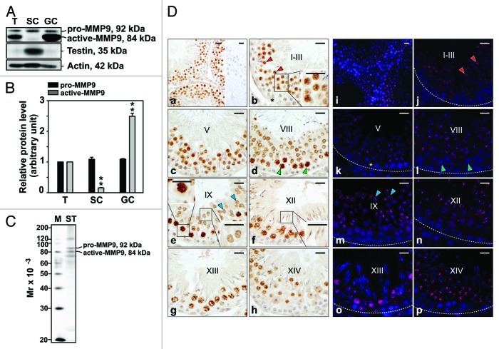
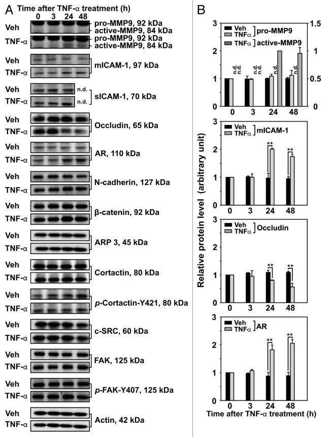
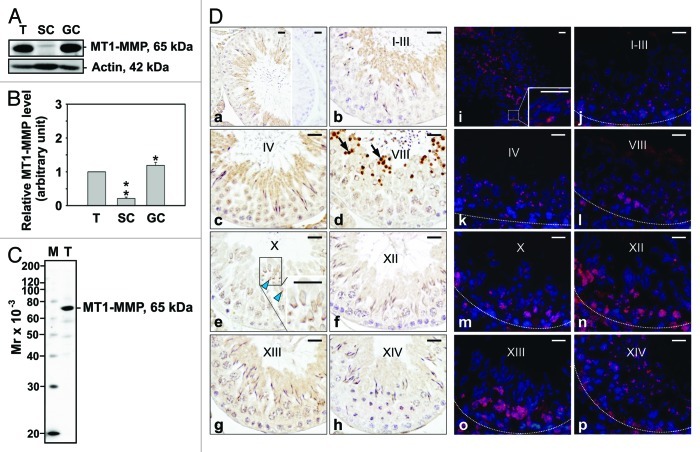
The mammalian blood-testis barrier (BTB) restructures throughout spermatogenesis, thereby allowing developing germ cells to enter the adluminal compartment of the seminiferous epithelium. Previous studies have shown pro-inflammatory cytokines such as tumor necrosis factor α (TNFα) and interleukin-1α to be important regulators of Sertoli cell barrier/BTB function in vitro and in vivo. In this study, the effects of TNFα on Sertoli cell barrier function were assessed, with emphasis on changes in proteases and cell adhesion molecules following treatment. By immunoblotting and immunohistochemistry, MMP9 was found to be present in germ cells, localizing by and large to spermatocytes and spermatids in the adult rat testis. Following treatment of Sertoli cells with physiologically relevant consecutive doses of recombinant human TNFα (25 ng/ml), the steady-state levels of active-matrix metalloprotease 9 (MMP9), membrane-bound intercellular adhesion molecule (mICAM-1) and androgen receptor increased significantly. TNFα also downregulated the steady-state level of occludin, in agreement with earlier results that showed TNFα to disrupt Sertoli cell barrier/BTB function. In addition, TNFα affected the filamentous actin cytoskeleton in Sertoli cells, which appeared to be mediated by cortactin, a regulator of actin dynamics. Taken collectively, these findings imply that germ cells may be involved in BTB restructuring via the localized production of TNFα. These results also illustrate that barrier restructuring correlated with an increase in Sertoli cell mICAM-1, suggesting that it may be critical for adhesion as germ cells traverse the "opened" BTB.

 求助内容:
求助内容: 应助结果提醒方式:


