Kok Chun Chang, Dino Samartzis, Keith D K Luk, Kenneth M C Cheung, Yat-Wa Wong
{"title":"Acute spinal subdural hematoma complicating lumbar decompressive surgery.","authors":"Kok Chun Chang, Dino Samartzis, Keith D K Luk, Kenneth M C Cheung, Yat-Wa Wong","doi":"10.1055/s-0031-1298602","DOIUrl":null,"url":null,"abstract":"<p><strong>Study design: </strong> A case report.</p><p><strong>Objective: </strong> To report a rare case of acute spinal subdural hematoma (SSH) complicating lumbar spine surgery, its characteristic presenting symptoms, diagnostic imaging, possible cause, and pitfall in management.</p><p><strong>Methods: </strong> A 59-year-old woman with lumbar spinal instability and stenosis underwent laminectomy and decompression at L3-L5 with instrumentation and fusion from L3-S1.</p><p><strong>Results: </strong> Immediately following surgery, the patient presented with incapacitating pain of both lower extremities from the mid-thigh downward, which was not relieved by narcotic analgesia and was disproportional to surgical trauma. Left ankle and great toes weakness was detected at postoperative day 2 and deteriorated on day 6. Magnetic resonance imaging was performed urgently and revealed a characteristic SSH with thecal sac compression at the level of L2, proximal to the laminectomy. Emergency decompression and evacuation of the hematoma was performed. The patient had partial recovery 6 weeks postoperatively.</p><p><strong>Conclusion: </strong> Acute SSH is a rare complication of lumbar spine surgery. This diagnosis must be considered when severe leg pain, unresolved with analgesia and disproportional to surgical trauma, with neurological deterioration occurring after lumbar spine surgery. Magnetic resonance imaging is the imaging modality of choice to assist in the differential diagnosis of an SSH. Early surgical decompression is necessary for optimal neurological recovery.</p>","PeriodicalId":89675,"journal":{"name":"Evidence-based spine-care journal","volume":"3 1","pages":"57-62"},"PeriodicalIF":0.0000,"publicationDate":"2012-02-01","publicationTypes":"Journal Article","fieldsOfStudy":null,"isOpenAccess":false,"openAccessPdf":"https://sci-hub-pdf.com/10.1055/s-0031-1298602","citationCount":"20","resultStr":null,"platform":"Semanticscholar","paperid":null,"PeriodicalName":"Evidence-based spine-care journal","FirstCategoryId":"1085","ListUrlMain":"https://doi.org/10.1055/s-0031-1298602","RegionNum":0,"RegionCategory":null,"ArticlePicture":[],"TitleCN":null,"AbstractTextCN":null,"PMCID":null,"EPubDate":"","PubModel":"","JCR":"","JCRName":"","Score":null,"Total":0}
引用次数: 20
Abstract
Study design: A case report.
Objective: To report a rare case of acute spinal subdural hematoma (SSH) complicating lumbar spine surgery, its characteristic presenting symptoms, diagnostic imaging, possible cause, and pitfall in management.
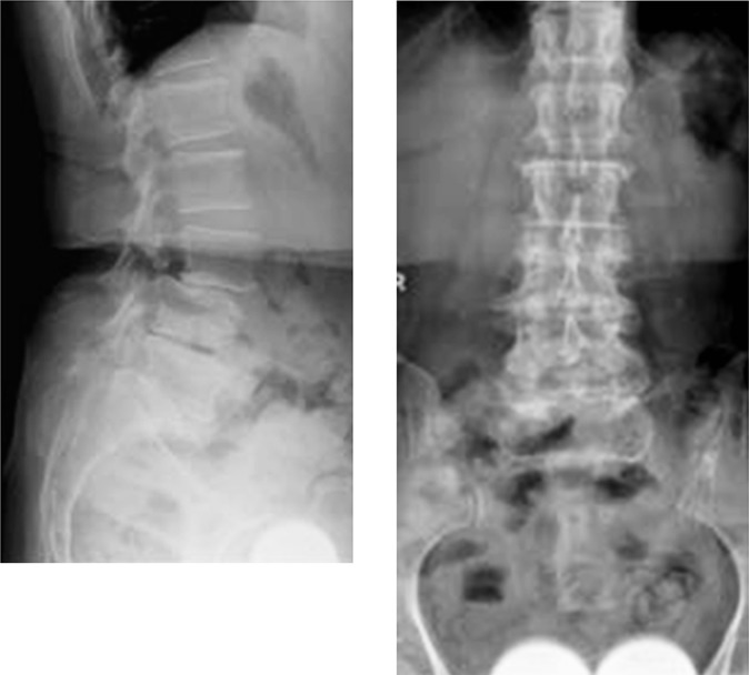
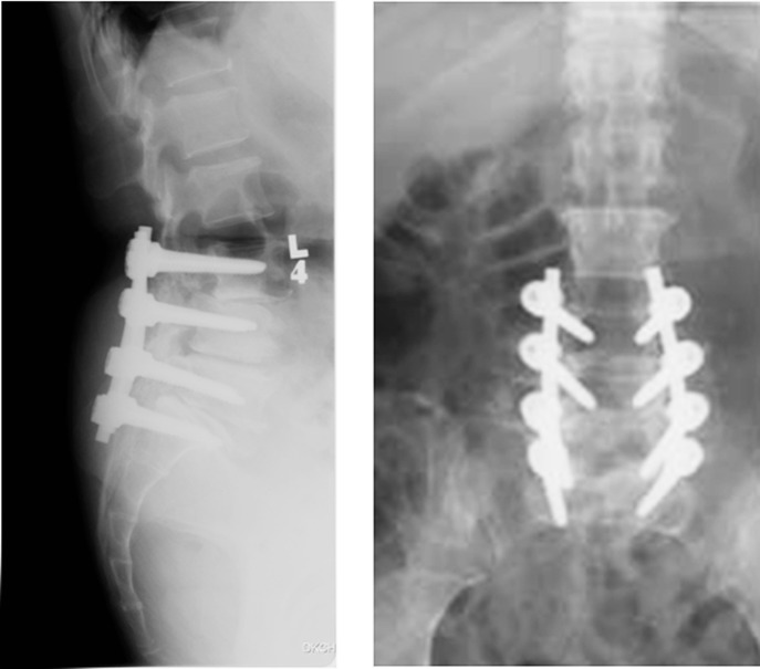
Methods: A 59-year-old woman with lumbar spinal instability and stenosis underwent laminectomy and decompression at L3-L5 with instrumentation and fusion from L3-S1.
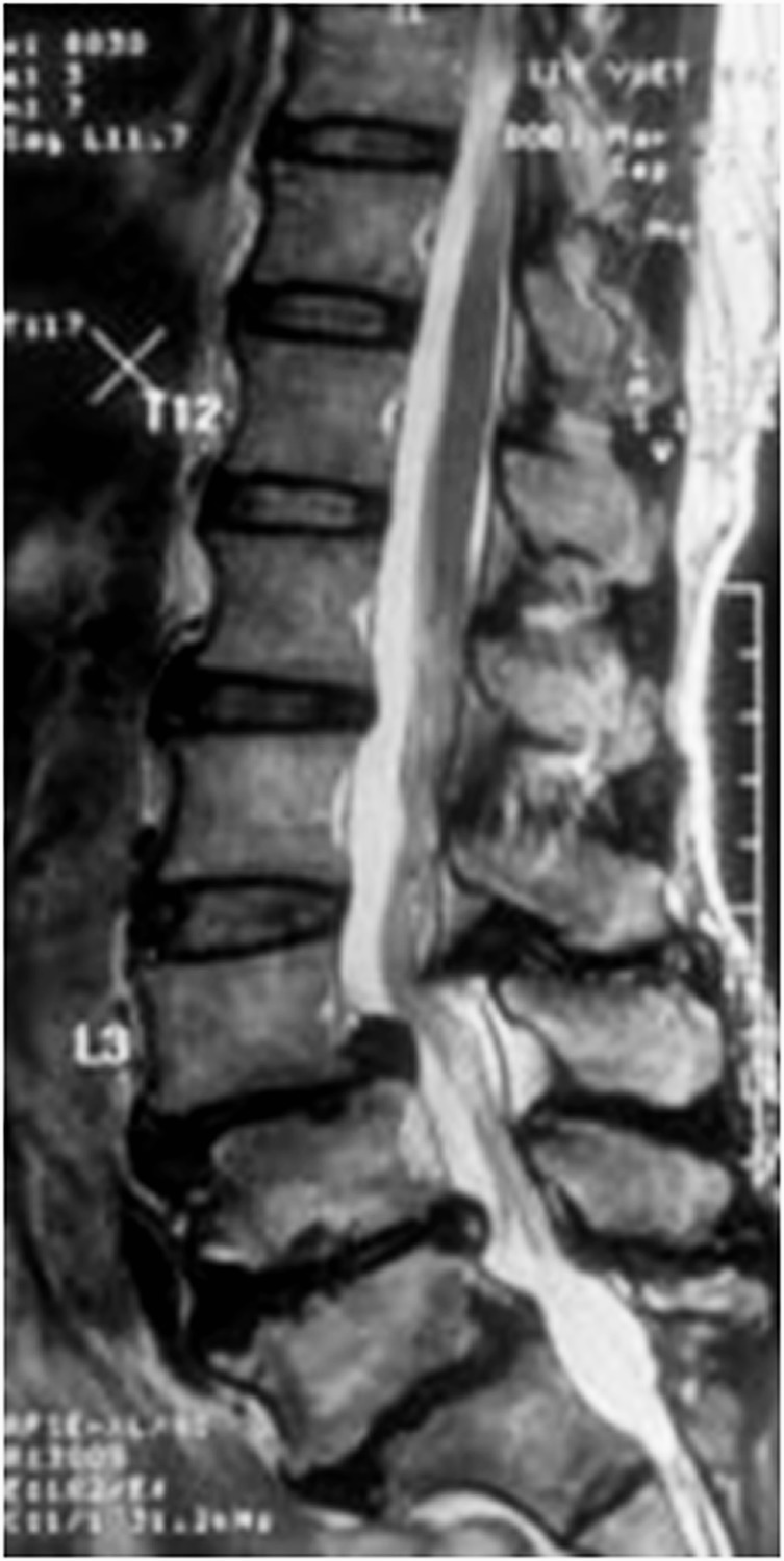
Results: Immediately following surgery, the patient presented with incapacitating pain of both lower extremities from the mid-thigh downward, which was not relieved by narcotic analgesia and was disproportional to surgical trauma. Left ankle and great toes weakness was detected at postoperative day 2 and deteriorated on day 6. Magnetic resonance imaging was performed urgently and revealed a characteristic SSH with thecal sac compression at the level of L2, proximal to the laminectomy. Emergency decompression and evacuation of the hematoma was performed. The patient had partial recovery 6 weeks postoperatively.
Conclusion: Acute SSH is a rare complication of lumbar spine surgery. This diagnosis must be considered when severe leg pain, unresolved with analgesia and disproportional to surgical trauma, with neurological deterioration occurring after lumbar spine surgery. Magnetic resonance imaging is the imaging modality of choice to assist in the differential diagnosis of an SSH. Early surgical decompression is necessary for optimal neurological recovery.

 求助内容:
求助内容: 应助结果提醒方式:


