{"title":"Arthroscopic resection of multiple ossifying tumors in the infrapatellar fat pad.","authors":"Tsutomu Oshigiri, Kota Watanabe, Hidenori Otsubo, Shintaro Takeda, Tomoyuki Suzuki, Takuma Kobayashi, Toshihiko Yamashita","doi":"10.1186/1758-2555-4-43","DOIUrl":null,"url":null,"abstract":"<p><p> A 49 year-old male visited a nearby clinic five years back with a complaint of pain in the right knee during exercise. Plain radiographs revealed absence of any anomalies. He began to feel a lumpy mass in his right knee two years back. The pain worsened, on imaging, an anomaly was identified in the infrapatellar fat pad of his right knee, and he was subsequently referred to our department where he was hospitalized. On examination, a mass extending on either side of the patellar tendon was identified along with rigid tenderness in that area. The knee's range of motion was 0degrees-130degrees, and knee flexion was accompanied by pain. The results of blood tests were normal. A plain radiograph of the knee revealed multiple ossifying tumors at a site consistent with the infrapatellar fat pad. T1-weighted MRI exhibited low-signal intensity, while T2-weighted MRI exhibited a mosaic-shaped tumor. We performed arthroscopic surgery to excise the tumor. The patient resumed work shortly after surgery and did not experience any pain during the two year postoperative observation period. The joint's range of motion improved to the extent that it was comparable with that of the left knee. No recurrence was observed on radiographic examination. In past studies, resection of similar tumors has been performed with an arthrotomy; however, we performed arthroscopic resection on our patient, who demonstrated a quick improvement in symptoms and range of motion after surgery. We believe that arthroscopic surgery is a feasible option to consider while treating such cases.</p>","PeriodicalId":88316,"journal":{"name":"Sports medicine, arthroscopy, rehabilitation, therapy & technology : SMARTT","volume":"4 1","pages":"43"},"PeriodicalIF":0.0000,"publicationDate":"2012-11-12","publicationTypes":"Journal Article","fieldsOfStudy":null,"isOpenAccess":false,"openAccessPdf":"https://sci-hub-pdf.com/10.1186/1758-2555-4-43","citationCount":"2","resultStr":null,"platform":"Semanticscholar","paperid":null,"PeriodicalName":"Sports medicine, arthroscopy, rehabilitation, therapy & technology : SMARTT","FirstCategoryId":"1085","ListUrlMain":"https://doi.org/10.1186/1758-2555-4-43","RegionNum":0,"RegionCategory":null,"ArticlePicture":[],"TitleCN":null,"AbstractTextCN":null,"PMCID":null,"EPubDate":"","PubModel":"","JCR":"","JCRName":"","Score":null,"Total":0}
引用次数: 2
Abstract
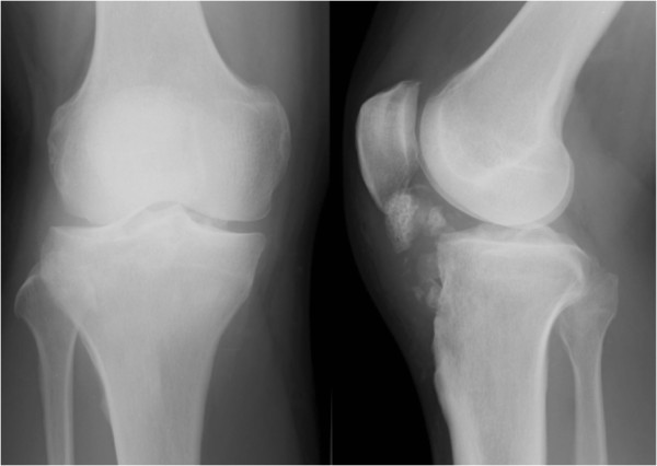
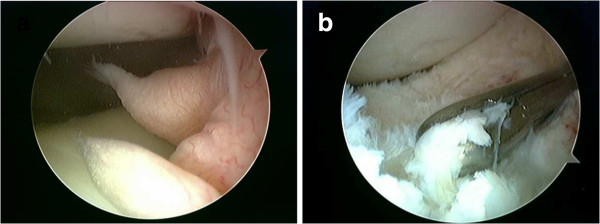
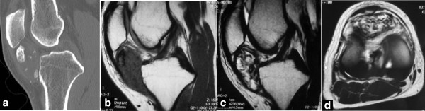
A 49 year-old male visited a nearby clinic five years back with a complaint of pain in the right knee during exercise. Plain radiographs revealed absence of any anomalies. He began to feel a lumpy mass in his right knee two years back. The pain worsened, on imaging, an anomaly was identified in the infrapatellar fat pad of his right knee, and he was subsequently referred to our department where he was hospitalized. On examination, a mass extending on either side of the patellar tendon was identified along with rigid tenderness in that area. The knee's range of motion was 0degrees-130degrees, and knee flexion was accompanied by pain. The results of blood tests were normal. A plain radiograph of the knee revealed multiple ossifying tumors at a site consistent with the infrapatellar fat pad. T1-weighted MRI exhibited low-signal intensity, while T2-weighted MRI exhibited a mosaic-shaped tumor. We performed arthroscopic surgery to excise the tumor. The patient resumed work shortly after surgery and did not experience any pain during the two year postoperative observation period. The joint's range of motion improved to the extent that it was comparable with that of the left knee. No recurrence was observed on radiographic examination. In past studies, resection of similar tumors has been performed with an arthrotomy; however, we performed arthroscopic resection on our patient, who demonstrated a quick improvement in symptoms and range of motion after surgery. We believe that arthroscopic surgery is a feasible option to consider while treating such cases.

 求助内容:
求助内容: 应助结果提醒方式:


