{"title":"Reversible conversion of epithelial and mesenchymal phenotypes in SV40 large T antigen-immortalized rat liver cell lines","authors":"Takato Takenouchi, Miyako Yoshioka, Noriko Yamanaka, Hiroshi Kitani","doi":"10.1042/CBR20100001","DOIUrl":null,"url":null,"abstract":"<p>EMT (epithelial—mesenchymal transition) is a key process in the development of liver fibrosis. This process is also essential for liver morphogenesis in embryonic development. To study the cellular and molecular basis of EMT, we established two phenotypically different SV40 large T antigen-immortalized cell lines from rat hepatocytes. The first cell line, which had an epithelial morphology and was established in DMEM (Dulbecco's modified Eagle's medium)/Ham's F-12 (DF)-based medium (RL/DF cells), expressed CK18 (cytokeratin 18), a marker of parenchymal hepatocytes. The other, a mesenchymal-like cell line established in DMEM-based medium (RL/DMEM cells), expressed αSMA (α-smooth muscle actin), a marker of hepatic myofibroblasts. Epithelial RL/DF cells underwent phenotypic changes, such as increased expression of αSMA, when the culture medium was switched to DMEM-based medium. In contrast, mesenchymal RL/DMEM cells were induced to express the epithelial marker CK18 with a concomitant decrease in αSMA expression when the culture medium was replaced with DF-based medium. These cell lines may provide novel <i>in vitro</i> models for the study of the conversion between epithelial and mesenchymal phenotypes during EMT in liver fibrosis and morphogenesis.</p>","PeriodicalId":75683,"journal":{"name":"Cell biology international reports","volume":"17 1","pages":"1-7"},"PeriodicalIF":0.0000,"publicationDate":"2013-06-25","publicationTypes":"Journal Article","fieldsOfStudy":null,"isOpenAccess":false,"openAccessPdf":"https://sci-hub-pdf.com/10.1042/CBR20100001","citationCount":"11","resultStr":null,"platform":"Semanticscholar","paperid":null,"PeriodicalName":"Cell biology international reports","FirstCategoryId":"1085","ListUrlMain":"https://onlinelibrary.wiley.com/doi/10.1042/CBR20100001","RegionNum":0,"RegionCategory":null,"ArticlePicture":[],"TitleCN":null,"AbstractTextCN":null,"PMCID":null,"EPubDate":"","PubModel":"","JCR":"","JCRName":"","Score":null,"Total":0}
引用次数: 11
Abstract
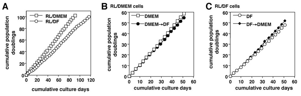
EMT (epithelial—mesenchymal transition) is a key process in the development of liver fibrosis. This process is also essential for liver morphogenesis in embryonic development. To study the cellular and molecular basis of EMT, we established two phenotypically different SV40 large T antigen-immortalized cell lines from rat hepatocytes. The first cell line, which had an epithelial morphology and was established in DMEM (Dulbecco's modified Eagle's medium)/Ham's F-12 (DF)-based medium (RL/DF cells), expressed CK18 (cytokeratin 18), a marker of parenchymal hepatocytes. The other, a mesenchymal-like cell line established in DMEM-based medium (RL/DMEM cells), expressed αSMA (α-smooth muscle actin), a marker of hepatic myofibroblasts. Epithelial RL/DF cells underwent phenotypic changes, such as increased expression of αSMA, when the culture medium was switched to DMEM-based medium. In contrast, mesenchymal RL/DMEM cells were induced to express the epithelial marker CK18 with a concomitant decrease in αSMA expression when the culture medium was replaced with DF-based medium. These cell lines may provide novel in vitro models for the study of the conversion between epithelial and mesenchymal phenotypes during EMT in liver fibrosis and morphogenesis.

 求助内容:
求助内容: 应助结果提醒方式:


