Jeffrey D Fineberg, David M Ritter, Manuel Covarrubias
{"title":"Modeling-independent elucidation of inactivation pathways in recombinant and native A-type Kv channels.","authors":"Jeffrey D Fineberg, David M Ritter, Manuel Covarrubias","doi":"10.1085/jgp.201210869","DOIUrl":null,"url":null,"abstract":"<p><p>A-type voltage-gated K(+) (Kv) channels self-regulate their activity by inactivating directly from the open state (open-state inactivation [OSI]) or by inactivating before they open (closed-state inactivation [CSI]). To determine the inactivation pathways, it is often necessary to apply several pulse protocols, pore blockers, single-channel recording, and kinetic modeling. However, intrinsic hurdles may preclude the standardized application of these methods. Here, we implemented a simple method inspired by earlier studies of Na(+) channels to analyze macroscopic inactivation and conclusively deduce the pathways of inactivation of recombinant and native A-type Kv channels. We investigated two distinct A-type Kv channels expressed heterologously (Kv3.4 and Kv4.2 with accessory subunits) and their native counterparts in dorsal root ganglion and cerebellar granule neurons. This approach applies two conventional pulse protocols to examine inactivation induced by (a) a simple step (single-pulse inactivation) and (b) a conditioning step (double-pulse inactivation). Consistent with OSI, the rate of Kv3.4 inactivation (i.e., the negative first derivative of double-pulse inactivation) precisely superimposes on the profile of the Kv3.4 current evoked by a single pulse because the channels must open to inactivate. In contrast, the rate of Kv4.2 inactivation is asynchronous, already changing at earlier times relative to the profile of the Kv4.2 current evoked by a single pulse. Thus, Kv4.2 inactivation occurs uncoupled from channel opening, indicating CSI. Furthermore, the inactivation time constant versus voltage relation of Kv3.4 decreases monotonically with depolarization and levels off, whereas that of Kv4.2 exhibits a J-shape profile. We also manipulated the inactivation phenotype by changing the subunit composition and show how CSI and CSI combined with OSI might affect spiking properties in a full computational model of the hippocampal CA1 neuron. This work unambiguously elucidates contrasting inactivation pathways in neuronal A-type Kv channels and demonstrates how distinct pathways might impact neurophysiological activity.</p>","PeriodicalId":173753,"journal":{"name":"The Journal of General Physiology","volume":" ","pages":"513-27"},"PeriodicalIF":0.0000,"publicationDate":"2012-11-01","publicationTypes":"Journal Article","fieldsOfStudy":null,"isOpenAccess":false,"openAccessPdf":"https://sci-hub-pdf.com/10.1085/jgp.201210869","citationCount":"25","resultStr":null,"platform":"Semanticscholar","paperid":null,"PeriodicalName":"The Journal of General Physiology","FirstCategoryId":"3","ListUrlMain":"https://doi.org/10.1085/jgp.201210869","RegionNum":0,"RegionCategory":null,"ArticlePicture":[],"TitleCN":null,"AbstractTextCN":null,"PMCID":null,"EPubDate":"","PubModel":"","JCR":"","JCRName":"","Score":null,"Total":0}
引用次数: 25
Abstract
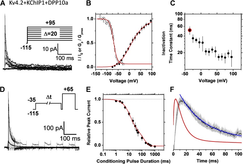
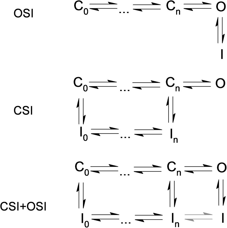
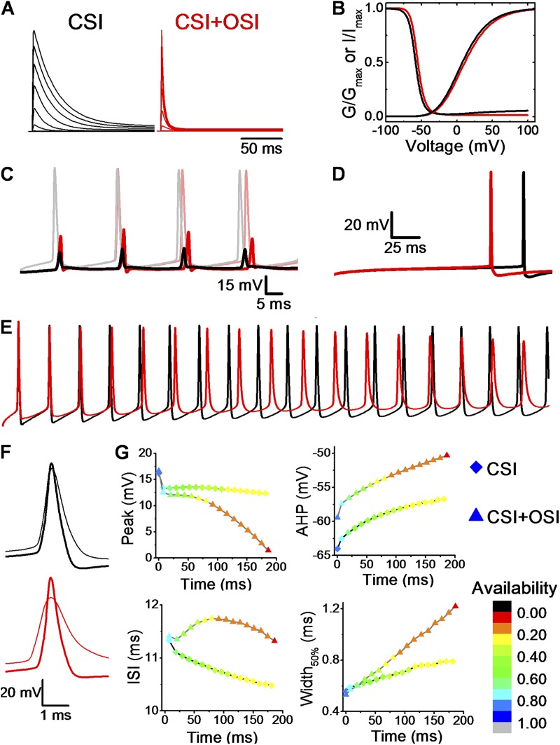
A-type voltage-gated K(+) (Kv) channels self-regulate their activity by inactivating directly from the open state (open-state inactivation [OSI]) or by inactivating before they open (closed-state inactivation [CSI]). To determine the inactivation pathways, it is often necessary to apply several pulse protocols, pore blockers, single-channel recording, and kinetic modeling. However, intrinsic hurdles may preclude the standardized application of these methods. Here, we implemented a simple method inspired by earlier studies of Na(+) channels to analyze macroscopic inactivation and conclusively deduce the pathways of inactivation of recombinant and native A-type Kv channels. We investigated two distinct A-type Kv channels expressed heterologously (Kv3.4 and Kv4.2 with accessory subunits) and their native counterparts in dorsal root ganglion and cerebellar granule neurons. This approach applies two conventional pulse protocols to examine inactivation induced by (a) a simple step (single-pulse inactivation) and (b) a conditioning step (double-pulse inactivation). Consistent with OSI, the rate of Kv3.4 inactivation (i.e., the negative first derivative of double-pulse inactivation) precisely superimposes on the profile of the Kv3.4 current evoked by a single pulse because the channels must open to inactivate. In contrast, the rate of Kv4.2 inactivation is asynchronous, already changing at earlier times relative to the profile of the Kv4.2 current evoked by a single pulse. Thus, Kv4.2 inactivation occurs uncoupled from channel opening, indicating CSI. Furthermore, the inactivation time constant versus voltage relation of Kv3.4 decreases monotonically with depolarization and levels off, whereas that of Kv4.2 exhibits a J-shape profile. We also manipulated the inactivation phenotype by changing the subunit composition and show how CSI and CSI combined with OSI might affect spiking properties in a full computational model of the hippocampal CA1 neuron. This work unambiguously elucidates contrasting inactivation pathways in neuronal A-type Kv channels and demonstrates how distinct pathways might impact neurophysiological activity.

 求助内容:
求助内容: 应助结果提醒方式:


