{"title":"Pelvic lipomatosis: US and CT diagnosis.","authors":"Y Yesilkaya, M Duymus, M Topcuoglu","doi":"10.2349/biij.8.2.e12","DOIUrl":null,"url":null,"abstract":"Pelvic lipomatosis is an uncommon benign disease that causes different symptoms due to the compression of pelvic organs by an intrapelvic overgrowth of mature fatty tissue. Engels first described the condition in 1959 [1]. The aetiology of the disease has not been established. However, it has been speculated that the fat proliferation might be associated with chronic pelvic inflammation due to chronic urinary tract infection [2]. In adults, endocrine diseases such as Cushing’s syndrome, hypothyroidism, insulin-secreting tumours, and neoplastic disease involving the hypothalamus (mainly craniopharyngioma) may also be a factor in the acquired obesity [2, 3]. Imaging is crucial in the diagnosis of pelvic lipomatosis. CT scans are considered to be the most effective and essential form of examination. The technique ensures dependable diagnosis of the disease principally because the absorption coefficient of the intrapelvic fatty tissue calculated by computer is distinct from that of other tissues. In the case we describe here, the diseases above could be ruled out based on the pattern of fat distribution, the clinical situation, and laboratory data. Laboratory results were all within normal limits. Pelvic lipomatosis typically exhibits a wide variety of symptoms including lumbago, discomfort of the lower abdomen, low-grade fever, recurrent urinary infections, frequent urination, dysuria, constipation, and hypertension [2, 4]. Our patient only had non-specific back pain.","PeriodicalId":89331,"journal":{"name":"Biomedical imaging and intervention journal","volume":"8 2","pages":"e12"},"PeriodicalIF":0.0000,"publicationDate":"2012-04-01","publicationTypes":"Journal Article","fieldsOfStudy":null,"isOpenAccess":false,"openAccessPdf":"https://sci-hub-pdf.com/10.2349/biij.8.2.e12","citationCount":"9","resultStr":null,"platform":"Semanticscholar","paperid":null,"PeriodicalName":"Biomedical imaging and intervention journal","FirstCategoryId":"1085","ListUrlMain":"https://doi.org/10.2349/biij.8.2.e12","RegionNum":0,"RegionCategory":null,"ArticlePicture":[],"TitleCN":null,"AbstractTextCN":null,"PMCID":null,"EPubDate":"","PubModel":"","JCR":"","JCRName":"","Score":null,"Total":0}
引用次数: 9
Abstract
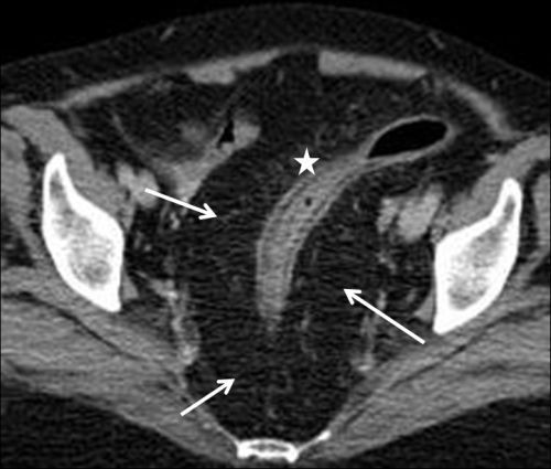
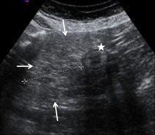
Pelvic lipomatosis is an uncommon benign disease that causes different symptoms due to the compression of pelvic organs by an intrapelvic overgrowth of mature fatty tissue. Engels first described the condition in 1959 [1]. The aetiology of the disease has not been established. However, it has been speculated that the fat proliferation might be associated with chronic pelvic inflammation due to chronic urinary tract infection [2]. In adults, endocrine diseases such as Cushing’s syndrome, hypothyroidism, insulin-secreting tumours, and neoplastic disease involving the hypothalamus (mainly craniopharyngioma) may also be a factor in the acquired obesity [2, 3]. Imaging is crucial in the diagnosis of pelvic lipomatosis. CT scans are considered to be the most effective and essential form of examination. The technique ensures dependable diagnosis of the disease principally because the absorption coefficient of the intrapelvic fatty tissue calculated by computer is distinct from that of other tissues. In the case we describe here, the diseases above could be ruled out based on the pattern of fat distribution, the clinical situation, and laboratory data. Laboratory results were all within normal limits. Pelvic lipomatosis typically exhibits a wide variety of symptoms including lumbago, discomfort of the lower abdomen, low-grade fever, recurrent urinary infections, frequent urination, dysuria, constipation, and hypertension [2, 4]. Our patient only had non-specific back pain.

 求助内容:
求助内容: 应助结果提醒方式:


