Honglan Wang, Ingrid M Bonilla, Xin Huang, Quanhua He, Mark J Kohr, Cynthia A Carnes, Mark T Ziolo
{"title":"Prolonged Action Potential and After depolarizations Are Not due to Changes in Potassium Currents in NOS3 Knockout Ventricular Myocytes.","authors":"Honglan Wang, Ingrid M Bonilla, Xin Huang, Quanhua He, Mark J Kohr, Cynthia A Carnes, Mark T Ziolo","doi":"10.1155/2012/645721","DOIUrl":null,"url":null,"abstract":"<p><p>Ventricular myocytes deficient in endothelial nitric oxide synthase (NOS3(-/-)) exhibit prolonged action potential (AP) duration and enhanced spontaneous activity (early and delayed afterdepolarizations) during β-adrenergic (β-AR) stimulation. Studies have shown that nitric oxide is able to regulate various K(+) channels. Our objective was to examine if NOS3(-/-) myocytes had altered K(+) currents. APs, transient outward (I(to)), sustained (I(Ksus)), and inward rectifier (I(K1)) K(+) currents were measured in NOS3(-/-) and wild-type (WT) myocytes. During β-AR stimulation, AP duration (measured as 90% repolarization-APD(90)) was prolonged in NOS3(-/-) compared to WT myocytes. Nevertheless, we did not observe differences in I(to), I(Ksus), or I(K1) between WT and NOS3(-/-) myocytes. Our previous work showed that NOS3(-/-) myocytes had a greater Ca(2+) influx via L-type Ca(2+) channels with β-AR stimulation. Thus, we measured β-AR-stimulated SR Ca(2+) load and found a greater increase in NOS3(-/-) versus WT myocytes. Hence, our data suggest that the prolonged AP in NOS3(-/-) myocytes is not due to changes in I(to), I(Ksus), or I(K1). Furthermore, the increase in spontaneous activity in NOS3(-/-) myocytes may be due to a greater increase in SR Ca(2+) load. This may have important implications for heart failure patients, where arrhythmias are increased and NOS3 expression is decreased.</p>","PeriodicalId":89176,"journal":{"name":"Journal of signal transduction","volume":"2012 ","pages":"645721"},"PeriodicalIF":0.0000,"publicationDate":"2012-01-01","publicationTypes":"Journal Article","fieldsOfStudy":null,"isOpenAccess":false,"openAccessPdf":"https://sci-hub-pdf.com/10.1155/2012/645721","citationCount":"10","resultStr":null,"platform":"Semanticscholar","paperid":null,"PeriodicalName":"Journal of signal transduction","FirstCategoryId":"1085","ListUrlMain":"https://doi.org/10.1155/2012/645721","RegionNum":0,"RegionCategory":null,"ArticlePicture":[],"TitleCN":null,"AbstractTextCN":null,"PMCID":null,"EPubDate":"2012/8/28 0:00:00","PubModel":"Epub","JCR":"","JCRName":"","Score":null,"Total":0}
引用次数: 10
Abstract
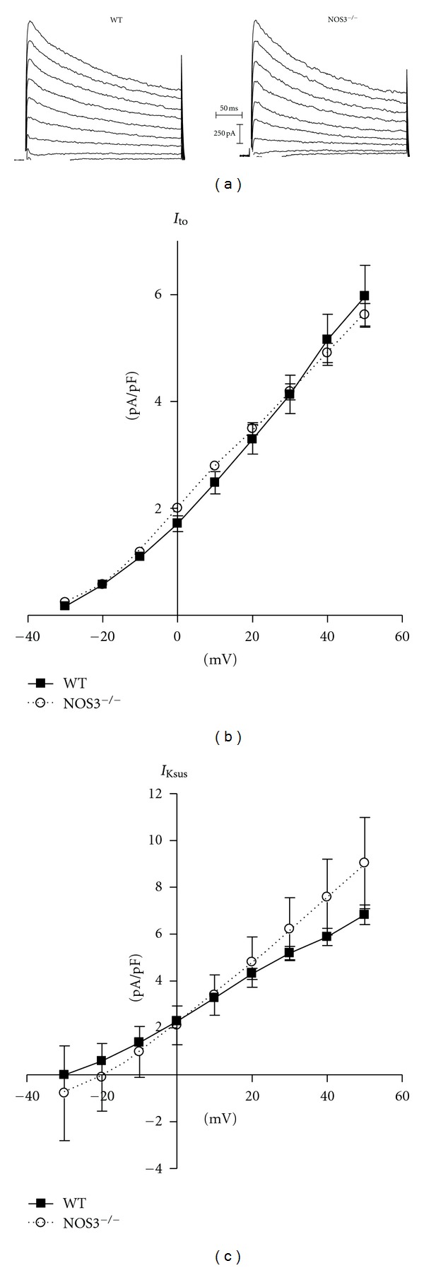
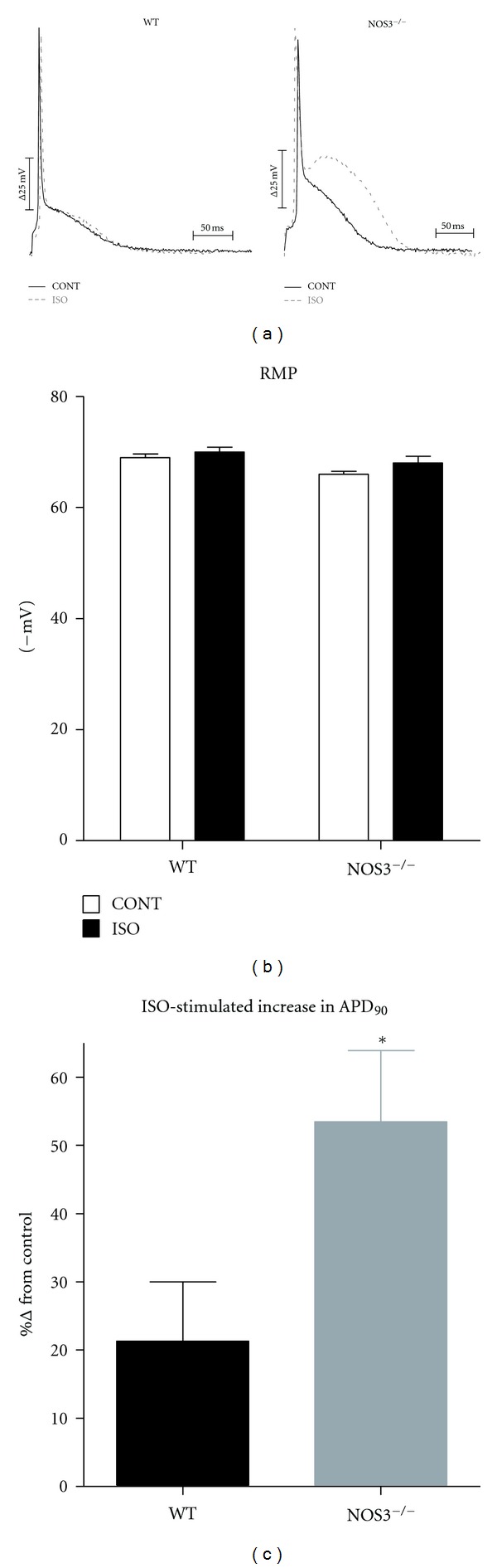
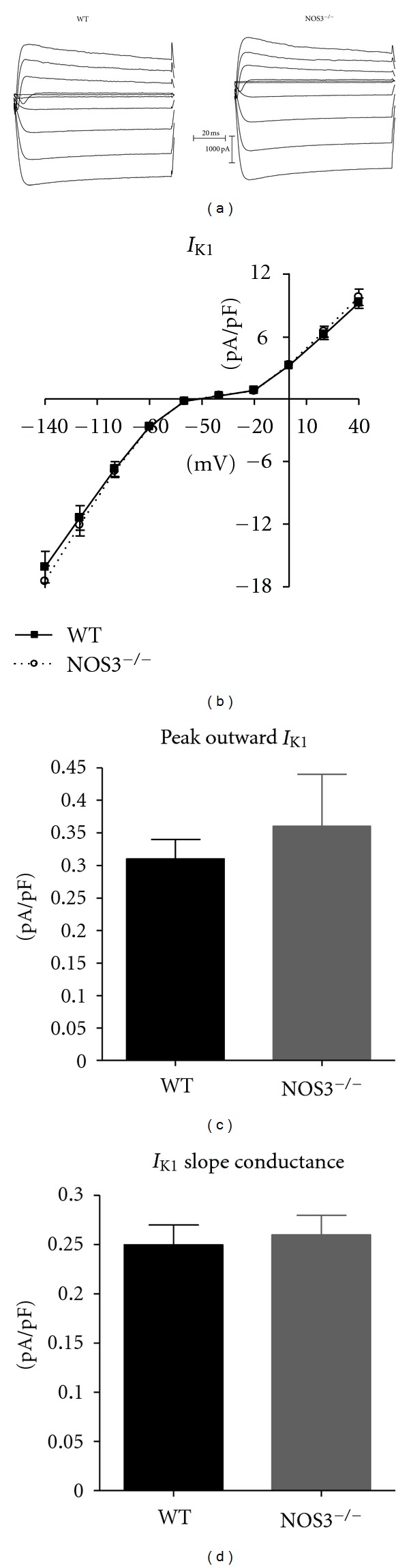
Ventricular myocytes deficient in endothelial nitric oxide synthase (NOS3(-/-)) exhibit prolonged action potential (AP) duration and enhanced spontaneous activity (early and delayed afterdepolarizations) during β-adrenergic (β-AR) stimulation. Studies have shown that nitric oxide is able to regulate various K(+) channels. Our objective was to examine if NOS3(-/-) myocytes had altered K(+) currents. APs, transient outward (I(to)), sustained (I(Ksus)), and inward rectifier (I(K1)) K(+) currents were measured in NOS3(-/-) and wild-type (WT) myocytes. During β-AR stimulation, AP duration (measured as 90% repolarization-APD(90)) was prolonged in NOS3(-/-) compared to WT myocytes. Nevertheless, we did not observe differences in I(to), I(Ksus), or I(K1) between WT and NOS3(-/-) myocytes. Our previous work showed that NOS3(-/-) myocytes had a greater Ca(2+) influx via L-type Ca(2+) channels with β-AR stimulation. Thus, we measured β-AR-stimulated SR Ca(2+) load and found a greater increase in NOS3(-/-) versus WT myocytes. Hence, our data suggest that the prolonged AP in NOS3(-/-) myocytes is not due to changes in I(to), I(Ksus), or I(K1). Furthermore, the increase in spontaneous activity in NOS3(-/-) myocytes may be due to a greater increase in SR Ca(2+) load. This may have important implications for heart failure patients, where arrhythmias are increased and NOS3 expression is decreased.

 求助内容:
求助内容: 应助结果提醒方式:


