Computed Tomography Evaluation of Craniomandibular Articulation in Class II Division 1 Malocclusion and Class I Normal Occlusion Subjects in North Indian Population.
K C Prabhat, Sanjeev Kumar Verma, Sandhya Maheshwari, Ibne Ahmad, Mohd Tariq
{"title":"Computed Tomography Evaluation of Craniomandibular Articulation in Class II Division 1 Malocclusion and Class I Normal Occlusion Subjects in North Indian Population.","authors":"K C Prabhat, Sanjeev Kumar Verma, Sandhya Maheshwari, Ibne Ahmad, Mohd Tariq","doi":"10.5402/2012/312031","DOIUrl":null,"url":null,"abstract":"<p><p>Objective. The purpose of this study is to investigate the Craniomandibular articulation morphology and position of condyle in mandibular fossae in Angle's class I normal occlusion and Angle's class II division 1 malocclusion. Materials and Methods. The present study was conducted on 40 subjects with 20 subjects in each group, and the computed tomography images were obtained using spiral computed tomography technique. Each measurement was compared by two-factor analysis of variance (ANOVA) while changes in anterior and posterior joint spaces were done by paired t-test. Results. Statistically significant anterior positioning of condyle (P > 0.05) was observed in class I normal malocclusion, and it was significant only on right side in class II division 1 malocclusion. Conclusions. There was no difference found in the condylar process and joint morphology between right and left sides of both Angle's Class I normal occlusion and Angle's class II division 1 malocclusion. Evaluation of the position of the condyles in their respective mandibular fossae showed concentric position with a tendency towards anterior positioning for both right and left sides of the subjects with Angle's Class I normal occlusion as well as subjects with Angle's class II division 1 malocclusion.</p>","PeriodicalId":89396,"journal":{"name":"ISRN dentistry","volume":"2012 ","pages":"312031"},"PeriodicalIF":0.0000,"publicationDate":"2012-01-01","publicationTypes":"Journal Article","fieldsOfStudy":null,"isOpenAccess":false,"openAccessPdf":"https://sci-hub-pdf.com/10.5402/2012/312031","citationCount":"7","resultStr":null,"platform":"Semanticscholar","paperid":null,"PeriodicalName":"ISRN dentistry","FirstCategoryId":"1085","ListUrlMain":"https://doi.org/10.5402/2012/312031","RegionNum":0,"RegionCategory":null,"ArticlePicture":[],"TitleCN":null,"AbstractTextCN":null,"PMCID":null,"EPubDate":"2012/8/16 0:00:00","PubModel":"Epub","JCR":"","JCRName":"","Score":null,"Total":0}
引用次数: 7
Abstract
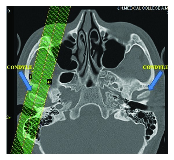
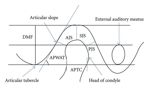
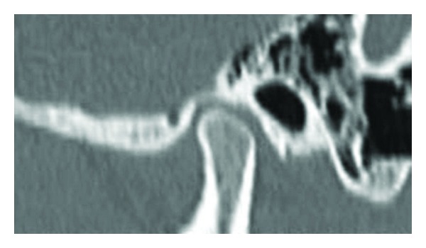
Objective. The purpose of this study is to investigate the Craniomandibular articulation morphology and position of condyle in mandibular fossae in Angle's class I normal occlusion and Angle's class II division 1 malocclusion. Materials and Methods. The present study was conducted on 40 subjects with 20 subjects in each group, and the computed tomography images were obtained using spiral computed tomography technique. Each measurement was compared by two-factor analysis of variance (ANOVA) while changes in anterior and posterior joint spaces were done by paired t-test. Results. Statistically significant anterior positioning of condyle (P > 0.05) was observed in class I normal malocclusion, and it was significant only on right side in class II division 1 malocclusion. Conclusions. There was no difference found in the condylar process and joint morphology between right and left sides of both Angle's Class I normal occlusion and Angle's class II division 1 malocclusion. Evaluation of the position of the condyles in their respective mandibular fossae showed concentric position with a tendency towards anterior positioning for both right and left sides of the subjects with Angle's Class I normal occlusion as well as subjects with Angle's class II division 1 malocclusion.

 求助内容:
求助内容: 应助结果提醒方式:


