{"title":"Anatomy of the temporal lobe.","authors":"J A Kiernan","doi":"10.1155/2012/176157","DOIUrl":null,"url":null,"abstract":"<p><p>Only primates have temporal lobes, which are largest in man, accommodating 17% of the cerebral cortex and including areas with auditory, olfactory, vestibular, visual and linguistic functions. The hippocampal formation, on the medial side of the lobe, includes the parahippocampal gyrus, subiculum, hippocampus, dentate gyrus, and associated white matter, notably the fimbria, whose fibres continue into the fornix. The hippocampus is an inrolled gyrus that bulges into the temporal horn of the lateral ventricle. Association fibres connect all parts of the cerebral cortex with the parahippocampal gyrus and subiculum, which in turn project to the dentate gyrus. The largest efferent projection of the subiculum and hippocampus is through the fornix to the hypothalamus. The choroid fissure, alongside the fimbria, separates the temporal lobe from the optic tract, hypothalamus and midbrain. The amygdala comprises several nuclei on the medial aspect of the temporal lobe, mostly anterior the hippocampus and indenting the tip of the temporal horn. The amygdala receives input from the olfactory bulb and from association cortex for other modalities of sensation. Its major projections are to the septal area and prefrontal cortex, mediating emotional responses to sensory stimuli. The temporal lobe contains much subcortical white matter, with such named bundles as the anterior commissure, arcuate fasciculus, inferior longitudinal fasciculus and uncinate fasciculus, and Meyer's loop of the geniculocalcarine tract. This article also reviews arterial supply, venous drainage, and anatomical relations of the temporal lobe to adjacent intracranial and tympanic structures.</p>","PeriodicalId":72948,"journal":{"name":"Epilepsy research and treatment","volume":" ","pages":"176157"},"PeriodicalIF":0.0000,"publicationDate":"2012-01-01","publicationTypes":"Journal Article","fieldsOfStudy":null,"isOpenAccess":false,"openAccessPdf":"https://sci-hub-pdf.com/10.1155/2012/176157","citationCount":"139","resultStr":null,"platform":"Semanticscholar","paperid":null,"PeriodicalName":"Epilepsy research and treatment","FirstCategoryId":"1085","ListUrlMain":"https://doi.org/10.1155/2012/176157","RegionNum":0,"RegionCategory":null,"ArticlePicture":[],"TitleCN":null,"AbstractTextCN":null,"PMCID":null,"EPubDate":"2012/3/29 0:00:00","PubModel":"Epub","JCR":"","JCRName":"","Score":null,"Total":0}
引用次数: 139
Abstract
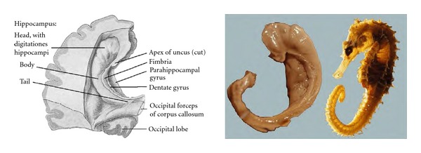
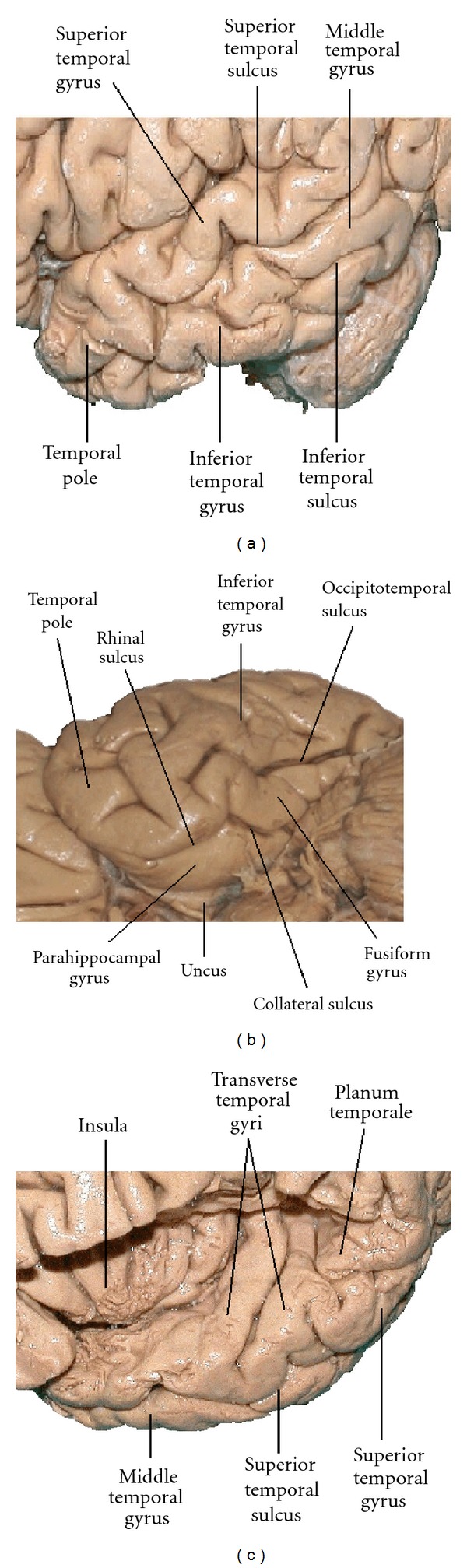
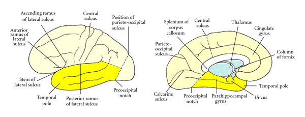
Only primates have temporal lobes, which are largest in man, accommodating 17% of the cerebral cortex and including areas with auditory, olfactory, vestibular, visual and linguistic functions. The hippocampal formation, on the medial side of the lobe, includes the parahippocampal gyrus, subiculum, hippocampus, dentate gyrus, and associated white matter, notably the fimbria, whose fibres continue into the fornix. The hippocampus is an inrolled gyrus that bulges into the temporal horn of the lateral ventricle. Association fibres connect all parts of the cerebral cortex with the parahippocampal gyrus and subiculum, which in turn project to the dentate gyrus. The largest efferent projection of the subiculum and hippocampus is through the fornix to the hypothalamus. The choroid fissure, alongside the fimbria, separates the temporal lobe from the optic tract, hypothalamus and midbrain. The amygdala comprises several nuclei on the medial aspect of the temporal lobe, mostly anterior the hippocampus and indenting the tip of the temporal horn. The amygdala receives input from the olfactory bulb and from association cortex for other modalities of sensation. Its major projections are to the septal area and prefrontal cortex, mediating emotional responses to sensory stimuli. The temporal lobe contains much subcortical white matter, with such named bundles as the anterior commissure, arcuate fasciculus, inferior longitudinal fasciculus and uncinate fasciculus, and Meyer's loop of the geniculocalcarine tract. This article also reviews arterial supply, venous drainage, and anatomical relations of the temporal lobe to adjacent intracranial and tympanic structures.

 求助内容:
求助内容: 应助结果提醒方式:


