Kevin Wang, Joanne H Wang, Harihara Baskaran, Russell Wang, Rick Jurevic
{"title":"Effect of human beta-defensin-3 on head and neck cancer cell migration using micro-fabricated cell islands.","authors":"Kevin Wang, Joanne H Wang, Harihara Baskaran, Russell Wang, Rick Jurevic","doi":"10.1186/1758-3284-4-41","DOIUrl":null,"url":null,"abstract":"<p><strong>Background: </strong>To examine the effect of the natural antimicrobial peptide human β-defensin-3 (hBD-3), on the migration of a head and neck cancer cell line in vitro using microfabrication and soft-lithographic techniques.</p><p><strong>Methods: </strong>TR146 cancer cells were seeded in Petri dishes with microfabricated wells for cell migration assays. Total 54 cell islands were used of various shape and size and experimental media type. Cell migration assays were analyzed in six group media: Dulbecco's modified medium (DMEM); DMEM with vascular endothelial growth factor (VEGF); Conditioned media of human embryonic kidney cells (HEK 239) expressing hBD-3 via transfected cloned pcDNA3 as CM/hBD-3; CM/hBD-3+ VEGF; conditioned medium from non-transfected HEK 239 (not expressing hBD-3) as control (CM); and the last group was CM + VEGF. Cell islands were circular or square and varied in size (0.25 mm(2), 0.125 mm(2), and 0.0625 mm(2)). Cell islands were imaged at t=0 h, 3 h, 6 h, and 24 h.</p><p><strong>Results: </strong>The results show cancer cell islands that originally were smaller had higher migration indices. There was no difference of MIs between circular and square cell islands. MIs at the end point were significantly different among the groups except between CM and CM-hBD-3+ VEGF.</p><p><strong>Conclusions: </strong>VEGF enhanced cancer cell migration. The combination of DMEM and VEGF showed a synergistic effect on this phenomenon of cancer cell migration. Conditioned medium with hBD-3 suppressed cancer cell migration. hBD-3 suppressed VEGF enhancement of TR146 cancer cell migration.</p>","PeriodicalId":49195,"journal":{"name":"Head and Neck Optical Diagnostics Society","volume":"4 ","pages":"41"},"PeriodicalIF":0.0000,"publicationDate":"2012-06-28","publicationTypes":"Journal Article","fieldsOfStudy":null,"isOpenAccess":false,"openAccessPdf":"https://sci-hub-pdf.com/10.1186/1758-3284-4-41","citationCount":"14","resultStr":null,"platform":"Semanticscholar","paperid":null,"PeriodicalName":"Head and Neck Optical Diagnostics Society","FirstCategoryId":"1085","ListUrlMain":"https://doi.org/10.1186/1758-3284-4-41","RegionNum":0,"RegionCategory":null,"ArticlePicture":[],"TitleCN":null,"AbstractTextCN":null,"PMCID":null,"EPubDate":"","PubModel":"","JCR":"","JCRName":"","Score":null,"Total":0}
引用次数: 14
Abstract
Background: To examine the effect of the natural antimicrobial peptide human β-defensin-3 (hBD-3), on the migration of a head and neck cancer cell line in vitro using microfabrication and soft-lithographic techniques.
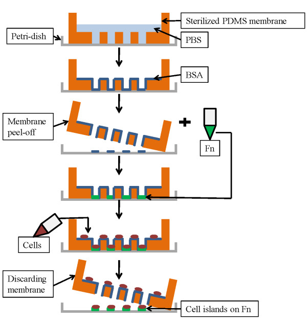
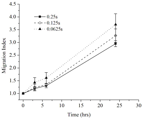
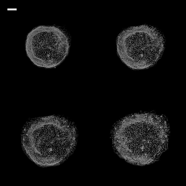
Methods: TR146 cancer cells were seeded in Petri dishes with microfabricated wells for cell migration assays. Total 54 cell islands were used of various shape and size and experimental media type. Cell migration assays were analyzed in six group media: Dulbecco's modified medium (DMEM); DMEM with vascular endothelial growth factor (VEGF); Conditioned media of human embryonic kidney cells (HEK 239) expressing hBD-3 via transfected cloned pcDNA3 as CM/hBD-3; CM/hBD-3+ VEGF; conditioned medium from non-transfected HEK 239 (not expressing hBD-3) as control (CM); and the last group was CM + VEGF. Cell islands were circular or square and varied in size (0.25 mm(2), 0.125 mm(2), and 0.0625 mm(2)). Cell islands were imaged at t=0 h, 3 h, 6 h, and 24 h.
Results: The results show cancer cell islands that originally were smaller had higher migration indices. There was no difference of MIs between circular and square cell islands. MIs at the end point were significantly different among the groups except between CM and CM-hBD-3+ VEGF.
Conclusions: VEGF enhanced cancer cell migration. The combination of DMEM and VEGF showed a synergistic effect on this phenomenon of cancer cell migration. Conditioned medium with hBD-3 suppressed cancer cell migration. hBD-3 suppressed VEGF enhancement of TR146 cancer cell migration.
 求助内容:
求助内容: 应助结果提醒方式:


