Zaid Hamdoon, Waseem Jerjes, Raed Al-Delayme, Colin Hopper
{"title":"Solitary giant neurofibroma of the neck subjected to photodynamic therapy: case study.","authors":"Zaid Hamdoon, Waseem Jerjes, Raed Al-Delayme, Colin Hopper","doi":"10.1186/1758-3284-4-30","DOIUrl":null,"url":null,"abstract":"<p><p>Photodynamic therapy (PDT) - the fourth modality - has been successfully used in the management of early and advanced pathologies of the head and neck. We studied the effect of this modality on a giant solitary neurofibroma of the neck. A 70-year-old Caucasian female presented with left neck pain and disfigurement associated with slight shortness of breath and dysphagia. Examination revealed a large mass in the neck with no neurovascular compromise. Magnetic resonance imaging (MRI) reported a heterogeneously enhancing mass extending from the left angle of the mandible to the base of the neck. A core biopsy was performed and histopathological examination revealed a disorganised array of peripheral nerve fascicles. The patient elected to receive photodynamic therapy as the primary intervention. The multi-disciplinary meeting approved the treatment plan. The photosensitizing agent was mTHPC (0.15 mg/kg), which was systemically administered 96-hours prior to ultrasound (US)-guided light delivery to the mass, which was undertaken under general anaesthesia. Recovery was uneventful. Post-PDT follow-up showed that the patient's pain, dysphagia and shortness of breath issues had improved. The disfigurement of the neck caused by the mass was no longer a problem. Three months post-PDT, MRI revealed a significant reduction in the neurofibroma size. PDT was proven as a successful primary intervention for this pathology. However, higher evidence-based studies are required before this therapy can be proposed as a replacement to any of the other conventional therapies.</p>","PeriodicalId":49195,"journal":{"name":"Head and Neck Optical Diagnostics Society","volume":"4 ","pages":"30"},"PeriodicalIF":0.0000,"publicationDate":"2012-01-01","publicationTypes":"Journal Article","fieldsOfStudy":null,"isOpenAccess":false,"openAccessPdf":"https://sci-hub-pdf.com/10.1186/1758-3284-4-30","citationCount":"17","resultStr":null,"platform":"Semanticscholar","paperid":null,"PeriodicalName":"Head and Neck Optical Diagnostics Society","FirstCategoryId":"1085","ListUrlMain":"https://doi.org/10.1186/1758-3284-4-30","RegionNum":0,"RegionCategory":null,"ArticlePicture":[],"TitleCN":null,"AbstractTextCN":null,"PMCID":null,"EPubDate":"2012/6/6 0:00:00","PubModel":"Epub","JCR":"","JCRName":"","Score":null,"Total":0}
引用次数: 17
Abstract
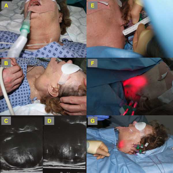
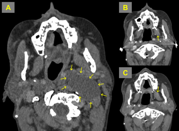
Photodynamic therapy (PDT) - the fourth modality - has been successfully used in the management of early and advanced pathologies of the head and neck. We studied the effect of this modality on a giant solitary neurofibroma of the neck. A 70-year-old Caucasian female presented with left neck pain and disfigurement associated with slight shortness of breath and dysphagia. Examination revealed a large mass in the neck with no neurovascular compromise. Magnetic resonance imaging (MRI) reported a heterogeneously enhancing mass extending from the left angle of the mandible to the base of the neck. A core biopsy was performed and histopathological examination revealed a disorganised array of peripheral nerve fascicles. The patient elected to receive photodynamic therapy as the primary intervention. The multi-disciplinary meeting approved the treatment plan. The photosensitizing agent was mTHPC (0.15 mg/kg), which was systemically administered 96-hours prior to ultrasound (US)-guided light delivery to the mass, which was undertaken under general anaesthesia. Recovery was uneventful. Post-PDT follow-up showed that the patient's pain, dysphagia and shortness of breath issues had improved. The disfigurement of the neck caused by the mass was no longer a problem. Three months post-PDT, MRI revealed a significant reduction in the neurofibroma size. PDT was proven as a successful primary intervention for this pathology. However, higher evidence-based studies are required before this therapy can be proposed as a replacement to any of the other conventional therapies.
 求助内容:
求助内容: 应助结果提醒方式:


