Se Heon Oh, Hwan Namgung, Mi Hyun Park, Dong-Guk Park
{"title":"Bezoar-induced Small Bowel Obstruction.","authors":"Se Heon Oh, Hwan Namgung, Mi Hyun Park, Dong-Guk Park","doi":"10.3393/jksc.2012.28.2.89","DOIUrl":null,"url":null,"abstract":"<p><strong>Purpose: </strong>The aim of this study was to observe the clinical features of a bezoar-induced small bowel obstruction and to investigate the role of abdominal computed tomography (CT) in establishing the diagnosis.</p><p><strong>Methods: </strong>We retrospectively reviewed 20 cases of bezoar-induced small bowel obstruction in our hospital from 1996 to 2010.</p><p><strong>Results: </strong>Thirteen patients (65%) had a history of abdominal surgery. Nine patients (45%) were diagnosed with a bezoar before surgery, seven patients were diagnosed by using abdominal CT, and two patients were diagnosed with a small bowel series. Abdominal CT was performed in 15 patients, and the diagnostic accuracy was 47% (7/15). Surgery revealed ten bezoars in the jejunum and 11 in the ileum. Two patients had bezoars found concurrently in the stomach. Spontaneous removal took place in two patients. An enterotomy and bezoar extraction was performed in 15 patients. Fragmentation and milking, a small bowel resection, and a Meckel's diverticulectomy were performed in one patient each. Early operative treatment was possible (P = 0.036) once the bezoar had been diagnosed by using abdominal CT. There tended to be fewer postoperative complications in patients who were diagnosed with a bezoar by using abdominal CT, but the result was not statistically significant (P = 0.712).</p><p><strong>Conclusion: </strong>A preoperative diagnosis of bezoar-induced small bowel obstruction by using clinical features was difficult. Increased use of abdominal CT led to a more accurate diagnosis and to earlier surgery for bezoar-induced small bowel obstructions, thereby reducing the rate of complications.</p>","PeriodicalId":17346,"journal":{"name":"Journal of the Korean Society of Coloproctology","volume":"28 2","pages":"89-93"},"PeriodicalIF":0.0000,"publicationDate":"2012-04-01","publicationTypes":"Journal Article","fieldsOfStudy":null,"isOpenAccess":false,"openAccessPdf":"https://ftp.ncbi.nlm.nih.gov/pub/pmc/oa_pdf/98/6f/jksc-28-89.PMC3349816.pdf","citationCount":"56","resultStr":null,"platform":"Semanticscholar","paperid":null,"PeriodicalName":"Journal of the Korean Society of Coloproctology","FirstCategoryId":"1085","ListUrlMain":"https://doi.org/10.3393/jksc.2012.28.2.89","RegionNum":0,"RegionCategory":null,"ArticlePicture":[],"TitleCN":null,"AbstractTextCN":null,"PMCID":null,"EPubDate":"2012/4/30 0:00:00","PubModel":"Epub","JCR":"","JCRName":"","Score":null,"Total":0}
引用次数: 56
Abstract
Purpose: The aim of this study was to observe the clinical features of a bezoar-induced small bowel obstruction and to investigate the role of abdominal computed tomography (CT) in establishing the diagnosis.
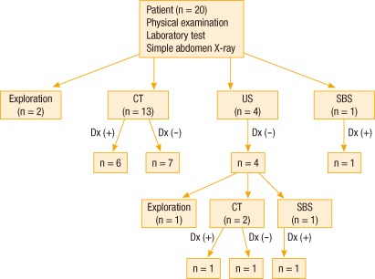
Methods: We retrospectively reviewed 20 cases of bezoar-induced small bowel obstruction in our hospital from 1996 to 2010.
Results: Thirteen patients (65%) had a history of abdominal surgery. Nine patients (45%) were diagnosed with a bezoar before surgery, seven patients were diagnosed by using abdominal CT, and two patients were diagnosed with a small bowel series. Abdominal CT was performed in 15 patients, and the diagnostic accuracy was 47% (7/15). Surgery revealed ten bezoars in the jejunum and 11 in the ileum. Two patients had bezoars found concurrently in the stomach. Spontaneous removal took place in two patients. An enterotomy and bezoar extraction was performed in 15 patients. Fragmentation and milking, a small bowel resection, and a Meckel's diverticulectomy were performed in one patient each. Early operative treatment was possible (P = 0.036) once the bezoar had been diagnosed by using abdominal CT. There tended to be fewer postoperative complications in patients who were diagnosed with a bezoar by using abdominal CT, but the result was not statistically significant (P = 0.712).
Conclusion: A preoperative diagnosis of bezoar-induced small bowel obstruction by using clinical features was difficult. Increased use of abdominal CT led to a more accurate diagnosis and to earlier surgery for bezoar-induced small bowel obstructions, thereby reducing the rate of complications.

 求助内容:
求助内容: 应助结果提醒方式:


