Joung Teak Jang, Haeng Ji Kang, Ji Young Yoon, Seo Gue Yoon
{"title":"Giant peritoneal loose body in the pelvic cavity.","authors":"Joung Teak Jang, Haeng Ji Kang, Ji Young Yoon, Seo Gue Yoon","doi":"10.3393/jksc.2012.28.2.108","DOIUrl":null,"url":null,"abstract":"<p><p>We report a case of a large peritoneal loose body diagnosed on computed tomography. The most common causes of a peritoneal loose body are thought to be torsion and separation of the appendices epiploicae. Peritoneal loose bodies are usually small, 0.5 to 2.5 cm in diameter. However, \"giant\" peritoneal loose bodies, larger than 4 cm in diameter, are an uncommon disease and present with various symptoms, and are difficult to diagnose preoperatively. Especially, abdominal large peritoneal loose bodies are frequently misdiagnosed as tumorous disease preoperatively. In our case, the loose body appeared as a round pelvic mass with central calcifications and a distinct fat plane separating it from adjacent organs. Preoperatively, we suspected a tumorous lesion from the wall of the upper rectum; however, at laparoscopy, a large peritoneal loose body was detected. An extraction of the giant peritoneal loose body was performed laparoscopically.</p>","PeriodicalId":17346,"journal":{"name":"Journal of the Korean Society of Coloproctology","volume":"28 2","pages":"108-10"},"PeriodicalIF":0.0000,"publicationDate":"2012-04-01","publicationTypes":"Journal Article","fieldsOfStudy":null,"isOpenAccess":false,"openAccessPdf":"https://ftp.ncbi.nlm.nih.gov/pub/pmc/oa_pdf/17/2c/jksc-28-108.PMC3349808.pdf","citationCount":"21","resultStr":null,"platform":"Semanticscholar","paperid":null,"PeriodicalName":"Journal of the Korean Society of Coloproctology","FirstCategoryId":"1085","ListUrlMain":"https://doi.org/10.3393/jksc.2012.28.2.108","RegionNum":0,"RegionCategory":null,"ArticlePicture":[],"TitleCN":null,"AbstractTextCN":null,"PMCID":null,"EPubDate":"2012/4/30 0:00:00","PubModel":"Epub","JCR":"","JCRName":"","Score":null,"Total":0}
引用次数: 21
Abstract
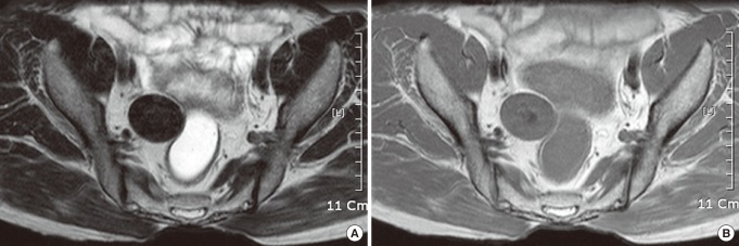
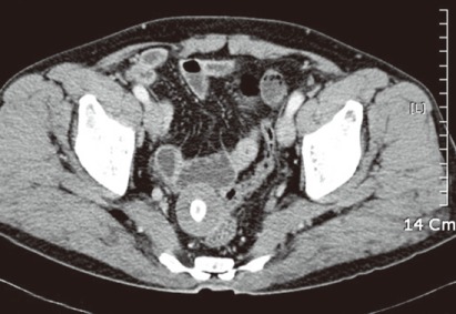
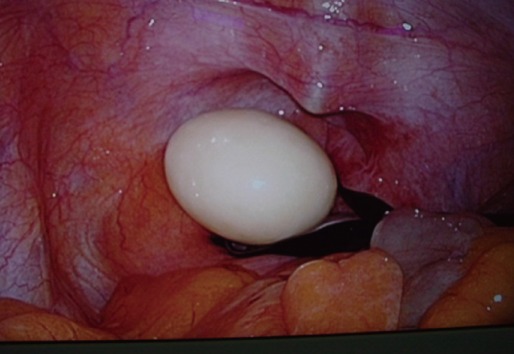
We report a case of a large peritoneal loose body diagnosed on computed tomography. The most common causes of a peritoneal loose body are thought to be torsion and separation of the appendices epiploicae. Peritoneal loose bodies are usually small, 0.5 to 2.5 cm in diameter. However, "giant" peritoneal loose bodies, larger than 4 cm in diameter, are an uncommon disease and present with various symptoms, and are difficult to diagnose preoperatively. Especially, abdominal large peritoneal loose bodies are frequently misdiagnosed as tumorous disease preoperatively. In our case, the loose body appeared as a round pelvic mass with central calcifications and a distinct fat plane separating it from adjacent organs. Preoperatively, we suspected a tumorous lesion from the wall of the upper rectum; however, at laparoscopy, a large peritoneal loose body was detected. An extraction of the giant peritoneal loose body was performed laparoscopically.

 求助内容:
求助内容: 应助结果提醒方式:


