Elizangela Partata Zuza, Ana Luiza Vanzato Carrareto, Raphael Carlos Comelli Lia, Juliana Rico Pires, Benedicto Egbert Corrêa de Toledo
{"title":"Histopathological features of dental pulp in teeth with different levels of chronic periodontitis severity.","authors":"Elizangela Partata Zuza, Ana Luiza Vanzato Carrareto, Raphael Carlos Comelli Lia, Juliana Rico Pires, Benedicto Egbert Corrêa de Toledo","doi":"10.5402/2012/271350","DOIUrl":null,"url":null,"abstract":"<p><p>Purpose. To evaluate the histopathological condition of the pulp in teeth with different levels of chronic periodontitis in humans. Methods. Twenty-five single-root nondecayed teeth were divided into three groups as follows: group 1, clinical attachment level (CAL) 3 to 4 mm and alveolar bone loss (BL) from 4 to 6 mm without reaching the tooth apex; group 2, CAL ≥ 5 mm and BL > 6 mm without reaching the tooth apex; group 3, CAL ≥ 5 mm and BL > 6 mm up to the tooth apex. Histological analyses were accomplished after laboratorial processing. Results. The mean of CAL was 3.2 ± 0.7 mm in group 1, 7.6 ± 2.0 mm in group 2, and 12.1 ± 2.8 mm in group 3, while for BL it was 4.8 ± 0.9 mm, 7.6 ± 2.2 mm, and 11.9 ± 2.1 mm, respectively. Histopathological data in the pulpal chambers were similar among the three groups showing normal aspects, and, the radicular pulps showed variable levels of reactive dentin, fibrosis, dystrophic mineralizations, atrophy, and mononuclear inflammatory infiltrate. Conclusions. Gradual progression of the chronic periodontitis led to changes in the histopathological aspects of the radicular pulp with progressive involvement.</p>","PeriodicalId":89396,"journal":{"name":"ISRN dentistry","volume":"2012 ","pages":"271350"},"PeriodicalIF":0.0000,"publicationDate":"2012-01-01","publicationTypes":"Journal Article","fieldsOfStudy":null,"isOpenAccess":false,"openAccessPdf":"https://sci-hub-pdf.com/10.5402/2012/271350","citationCount":"23","resultStr":null,"platform":"Semanticscholar","paperid":null,"PeriodicalName":"ISRN dentistry","FirstCategoryId":"1085","ListUrlMain":"https://doi.org/10.5402/2012/271350","RegionNum":0,"RegionCategory":null,"ArticlePicture":[],"TitleCN":null,"AbstractTextCN":null,"PMCID":null,"EPubDate":"2012/4/10 0:00:00","PubModel":"Epub","JCR":"","JCRName":"","Score":null,"Total":0}
引用次数: 23
Abstract
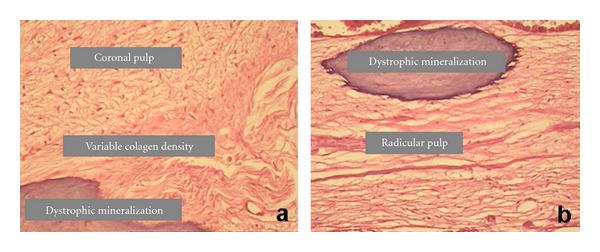
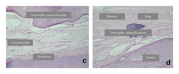
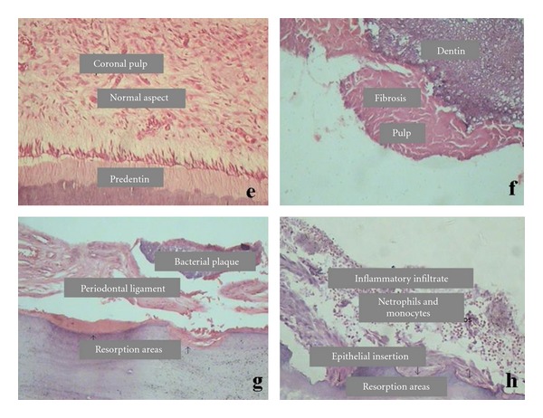
Purpose. To evaluate the histopathological condition of the pulp in teeth with different levels of chronic periodontitis in humans. Methods. Twenty-five single-root nondecayed teeth were divided into three groups as follows: group 1, clinical attachment level (CAL) 3 to 4 mm and alveolar bone loss (BL) from 4 to 6 mm without reaching the tooth apex; group 2, CAL ≥ 5 mm and BL > 6 mm without reaching the tooth apex; group 3, CAL ≥ 5 mm and BL > 6 mm up to the tooth apex. Histological analyses were accomplished after laboratorial processing. Results. The mean of CAL was 3.2 ± 0.7 mm in group 1, 7.6 ± 2.0 mm in group 2, and 12.1 ± 2.8 mm in group 3, while for BL it was 4.8 ± 0.9 mm, 7.6 ± 2.2 mm, and 11.9 ± 2.1 mm, respectively. Histopathological data in the pulpal chambers were similar among the three groups showing normal aspects, and, the radicular pulps showed variable levels of reactive dentin, fibrosis, dystrophic mineralizations, atrophy, and mononuclear inflammatory infiltrate. Conclusions. Gradual progression of the chronic periodontitis led to changes in the histopathological aspects of the radicular pulp with progressive involvement.
目的。探讨不同程度慢性牙周炎患者牙髓的组织病理学状况。方法。25颗单根无龋牙分为3组:1组临床附着水平(CAL)为3 ~ 4mm,牙槽骨损失(BL)为4 ~ 6mm,未到达牙尖;第2组,CAL≥5mm, BL > 6mm,但未到达牙尖;第3组,CAL≥5mm, BL > 6mm,直至齿尖。实验室处理后进行组织学分析。结果。1组CAL的平均值为3.2±0.7 mm, 2组为7.6±2.0 mm, 3组为12.1±2.8 mm, BL的平均值分别为4.8±0.9 mm, 7.6±2.2 mm, 11.9±2.1 mm。三组牙髓腔的组织病理学数据相似,显示正常,根状牙髓显示不同程度的反应性牙本质、纤维化、营养不良矿化、萎缩和单核炎症浸润。结论。慢性牙周炎的逐渐发展导致根状牙髓的组织病理学改变,并逐渐受累。

 求助内容:
求助内容: 应助结果提醒方式:


