Pediatric cerebellar hemorrhagic glioblastoma multiforme.

Q4 Medicine
Open Neuroimaging Journal Pub Date : 2012-01-01 Epub Date: 2012-02-28 DOI:10.2174/1874440001206010013
Peter Kalina
{"title":"Pediatric cerebellar hemorrhagic glioblastoma multiforme.","authors":"Peter Kalina","doi":"10.2174/1874440001206010013","DOIUrl":null,"url":null,"abstract":"<p><p>We report the case of an 11 year old boy who presented with nausea, vomiting and ataxia. He was evaluated with computed tomography (CT) and magnetic resonance imaging (MRI). Imaging demonstrated minimal enhancement and hemorrhage of a cerebellar mass. Cerebellar glioblastoma multiforme (GBM) is extremely rare in the cerebellum at any age but especially in children. The atypical findings of minimal enhancement, cerebellar location and hemorrhagic presentation combine to make the prospective diagnosis of GBM a difficult one. This rare combination of findings has not been previously reported.</p>","PeriodicalId":37431,"journal":{"name":"Open Neuroimaging Journal","volume":null,"pages":null},"PeriodicalIF":0.0000,"publicationDate":"2012-01-01","publicationTypes":"Journal Article","fieldsOfStudy":null,"isOpenAccess":false,"openAccessPdf":"https://ftp.ncbi.nlm.nih.gov/pub/pmc/oa_pdf/c9/4f/TONIJ-6-13.PMC3296109.pdf","citationCount":"5","resultStr":null,"platform":"Semanticscholar","paperid":null,"PeriodicalName":"Open Neuroimaging Journal","FirstCategoryId":"1085","ListUrlMain":"https://doi.org/10.2174/1874440001206010013","RegionNum":0,"RegionCategory":null,"ArticlePicture":[],"TitleCN":null,"AbstractTextCN":null,"PMCID":null,"EPubDate":"2012/2/28 0:00:00","PubModel":"Epub","JCR":"Q4","JCRName":"Medicine","Score":null,"Total":0}
引用次数: 5

Abstract

We report the case of an 11 year old boy who presented with nausea, vomiting and ataxia. He was evaluated with computed tomography (CT) and magnetic resonance imaging (MRI). Imaging demonstrated minimal enhancement and hemorrhage of a cerebellar mass. Cerebellar glioblastoma multiforme (GBM) is extremely rare in the cerebellum at any age but especially in children. The atypical findings of minimal enhancement, cerebellar location and hemorrhagic presentation combine to make the prospective diagnosis of GBM a difficult one. This rare combination of findings has not been previously reported.
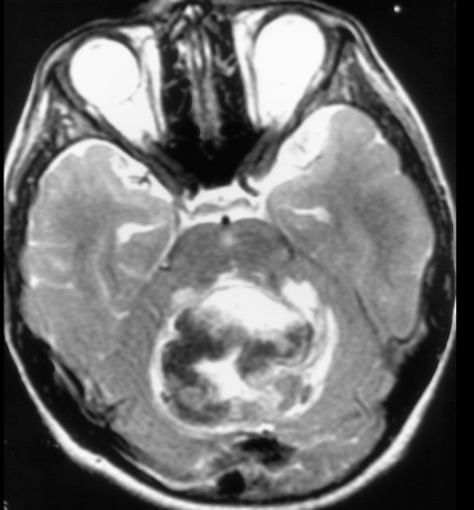
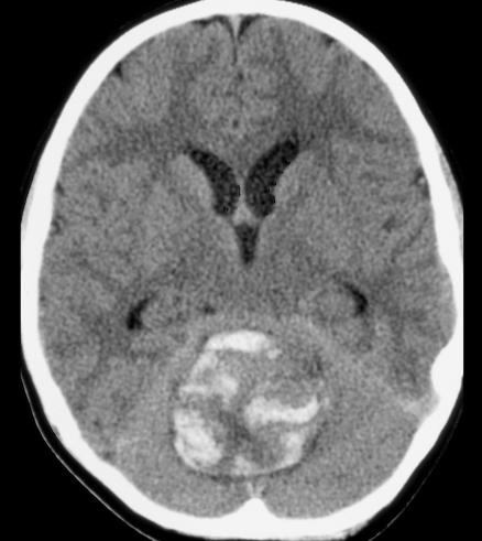
Abstract Image

Abstract Image

小儿多形性颅内出血性胶质母细胞瘤。
我们报告的情况下,11岁的男孩谁提出恶心,呕吐和共济失调。行计算机断层扫描(CT)和磁共振成像(MRI)检查。影像显示轻微强化及小脑肿块出血。多形性小脑胶质母细胞瘤(GBM)在任何年龄的小脑中都是极其罕见的,尤其是在儿童中。微小强化的不典型表现,小脑的位置和出血表现结合起来,使GBM的前瞻性诊断变得困难。这种罕见的组合发现以前没有报道过。
本文章由计算机程序翻译,如有差异,请以英文原文为准。
求助全文
约1分钟内获得全文 求助全文
来源期刊
Open Neuroimaging Journal
Open Neuroimaging Journal Medicine-Radiology, Nuclear Medicine and Imaging
CiteScore
0.70
自引率
0.00%
发文量
3
期刊介绍: The Open Neuroimaging Journal is an Open Access online journal, which publishes research articles, reviews/mini-reviews, and letters in all important areas of brain function, structure and organization including neuroimaging, neuroradiology, analysis methods, functional MRI acquisition and physics, brain mapping, macroscopic level of brain organization, computational modeling and analysis, structure-function and brain-behavior relationships, anatomy and physiology, psychiatric diseases and disorders of the nervous system, use of imaging to the understanding of brain pathology and brain abnormalities, cognition and aging, social neuroscience, sensorimotor processing, communication and learning.
×
引用
GB/T 7714-2015
复制
MLA
复制
APA
复制
导出至
BibTeX EndNote RefMan NoteFirst NoteExpress
×
提示
您的信息不完整,为了账户安全,请先补充。
现在去补充
×
提示
您因"违规操作"
具体请查看互助需知
我知道了
×
提示
确定
请完成安全验证×
copy
已复制链接
快去分享给好友吧!
我知道了
右上角分享
点击右上角分享
0
联系我们:info@booksci.cn Book学术提供免费学术资源搜索服务,方便国内外学者检索中英文文献。致力于提供最便捷和优质的服务体验。 Copyright © 2023 布克学术 All rights reserved.
京ICP备2023020795号-1
ghs 京公网安备 11010802042870号
Book学术文献互助
Book学术文献互助群
群 号:481959085
Book学术官方微信