{"title":"Tinea corporis gladiatorum presenting as a majocchi granuloma.","authors":"Anil Kurian, Richard M Haber","doi":"10.5402/2011/767589","DOIUrl":null,"url":null,"abstract":"<p><p>Background. Wrestlers are at increased risk of developing cutaneous infections, including fungal infections caused by dermatophytes. Erythematous lesions due to tinea infections can be mistakenly diagnosed as an inflammatory dermatitis and incorrectly treated with potent topical corticosteroid treatments which cause localized skin immunosuppression. This can eventuate in a Majocchi granuloma which then becomes refractory to topical antifungal therapy. To our knowledge, this is the first case of tinea corporis gladiatorum presenting as a Majocchi granuloma. Observations. A 20-year-old wrestler presented with a 4-year history of a large pruritic, scaly erythematous plaque with follicular papules, and pustules on his right forearm. The lesion had the clinical appearance of a Majocchi granuloma. He had been treated with potent topical corticosteroids and topical antifungal therapy. KOH and fungal culture of the lesion were negative. An erythematous scaly lesion in the scalp was cultured and grew Trichophyton tonsurans. Oral Terbinafine therapy was initiated and complete resolution of both lesions occurred within 6 weeks. Conclusion. The purpose of this report is to inform dermatologists that tinea corporis gladiatorum can present as a Majocchi granuloma and needs to be considered in the differential diagnosis of persistent skin lesions in wrestlers.</p>","PeriodicalId":14682,"journal":{"name":"ISRN Dermatology","volume":"2011 ","pages":"767589"},"PeriodicalIF":0.0000,"publicationDate":"2011-01-01","publicationTypes":"Journal Article","fieldsOfStudy":null,"isOpenAccess":false,"openAccessPdf":"https://sci-hub-pdf.com/10.5402/2011/767589","citationCount":"19","resultStr":null,"platform":"Semanticscholar","paperid":null,"PeriodicalName":"ISRN Dermatology","FirstCategoryId":"1085","ListUrlMain":"https://doi.org/10.5402/2011/767589","RegionNum":0,"RegionCategory":null,"ArticlePicture":[],"TitleCN":null,"AbstractTextCN":null,"PMCID":null,"EPubDate":"2011/4/12 0:00:00","PubModel":"Epub","JCR":"","JCRName":"","Score":null,"Total":0}
引用次数: 19
Abstract
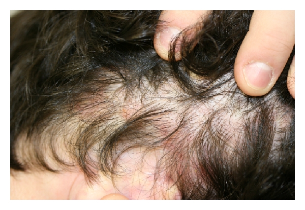
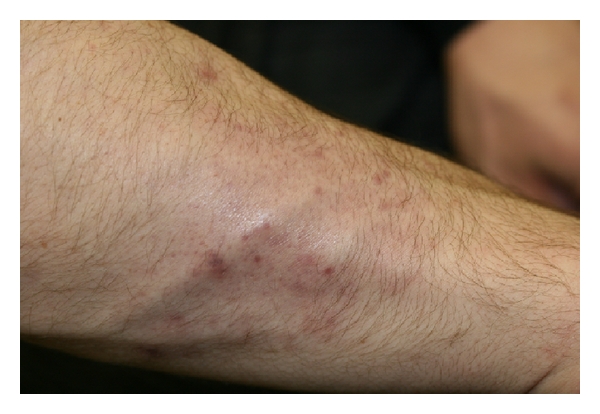
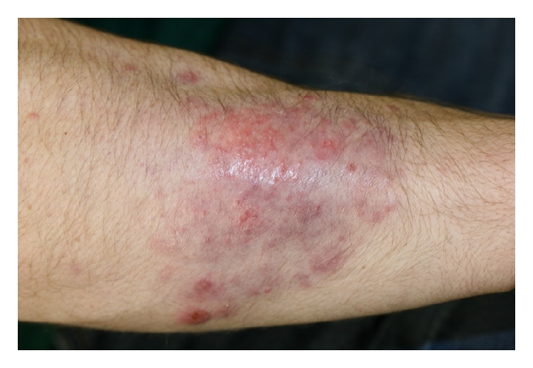
Background. Wrestlers are at increased risk of developing cutaneous infections, including fungal infections caused by dermatophytes. Erythematous lesions due to tinea infections can be mistakenly diagnosed as an inflammatory dermatitis and incorrectly treated with potent topical corticosteroid treatments which cause localized skin immunosuppression. This can eventuate in a Majocchi granuloma which then becomes refractory to topical antifungal therapy. To our knowledge, this is the first case of tinea corporis gladiatorum presenting as a Majocchi granuloma. Observations. A 20-year-old wrestler presented with a 4-year history of a large pruritic, scaly erythematous plaque with follicular papules, and pustules on his right forearm. The lesion had the clinical appearance of a Majocchi granuloma. He had been treated with potent topical corticosteroids and topical antifungal therapy. KOH and fungal culture of the lesion were negative. An erythematous scaly lesion in the scalp was cultured and grew Trichophyton tonsurans. Oral Terbinafine therapy was initiated and complete resolution of both lesions occurred within 6 weeks. Conclusion. The purpose of this report is to inform dermatologists that tinea corporis gladiatorum can present as a Majocchi granuloma and needs to be considered in the differential diagnosis of persistent skin lesions in wrestlers.

 求助内容:
求助内容: 应助结果提醒方式:


