Chun-Yang Wang, Jui-Che Tsai, Ching-Cheng Chuang, Yao-Sheng Hsieh, Chia-Wei Sun
{"title":"Aorta fluorescence imaging by using confocal microscopy.","authors":"Chun-Yang Wang, Jui-Che Tsai, Ching-Cheng Chuang, Yao-Sheng Hsieh, Chia-Wei Sun","doi":"10.5402/2011/215627","DOIUrl":null,"url":null,"abstract":"<p><p>The activated leukocyte attacked the vascular endothelium and the associated increase in VEcadherin number was observed in experiments. The confocal microscopic system with a prism-based wavelength filter was used for multiwavelength fluorescence measurement. Multiwavelength fluorescence imaging based on the VEcadherin within the aorta segment of a rat was achieved. The confocal microscopic system capable of fluorescence detection of cardiovascular tissue is a useful tool for measuring the biological properties in clinical applications.</p>","PeriodicalId":73519,"journal":{"name":"ISRN cardiology","volume":"2011 ","pages":"215627"},"PeriodicalIF":0.0000,"publicationDate":"2011-01-01","publicationTypes":"Journal Article","fieldsOfStudy":null,"isOpenAccess":false,"openAccessPdf":"https://sci-hub-pdf.com/10.5402/2011/215627","citationCount":"3","resultStr":null,"platform":"Semanticscholar","paperid":null,"PeriodicalName":"ISRN cardiology","FirstCategoryId":"1085","ListUrlMain":"https://doi.org/10.5402/2011/215627","RegionNum":0,"RegionCategory":null,"ArticlePicture":[],"TitleCN":null,"AbstractTextCN":null,"PMCID":null,"EPubDate":"2011/7/9 0:00:00","PubModel":"Epub","JCR":"","JCRName":"","Score":null,"Total":0}
引用次数: 3
Abstract
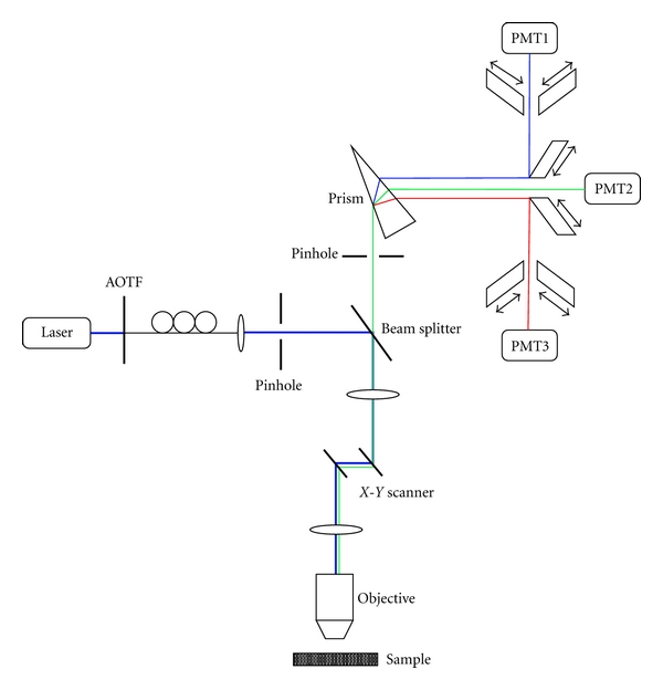
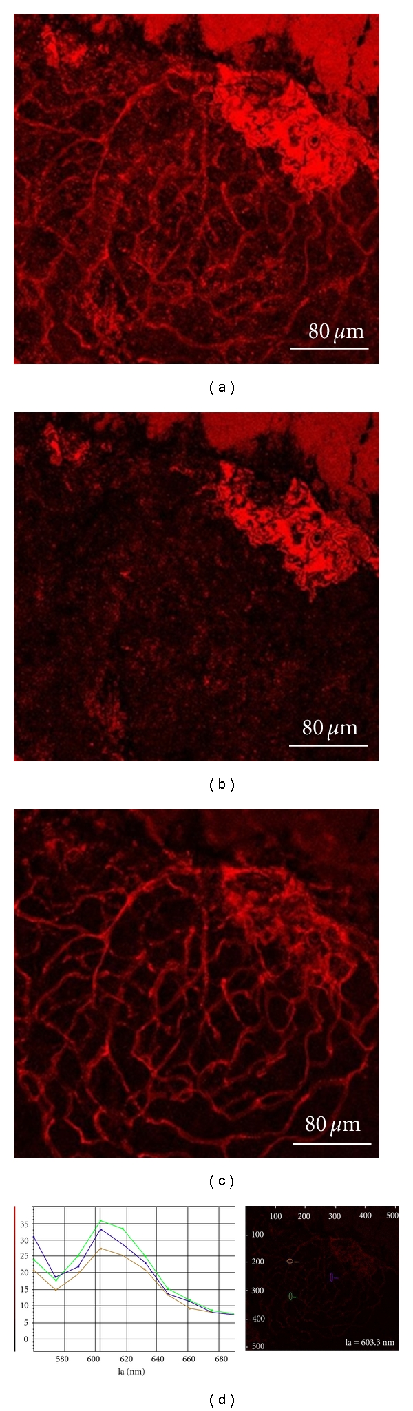
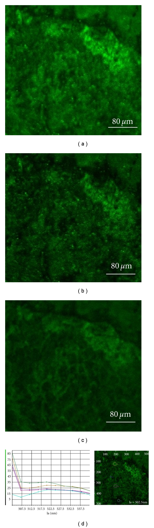
The activated leukocyte attacked the vascular endothelium and the associated increase in VEcadherin number was observed in experiments. The confocal microscopic system with a prism-based wavelength filter was used for multiwavelength fluorescence measurement. Multiwavelength fluorescence imaging based on the VEcadherin within the aorta segment of a rat was achieved. The confocal microscopic system capable of fluorescence detection of cardiovascular tissue is a useful tool for measuring the biological properties in clinical applications.

 求助内容:
求助内容: 应助结果提醒方式:


