Masanori Matsuzaki, Graham Cr Ellis-Davies, Yuya Kanemoto, Haruo Kasai
{"title":"Simultaneous two-photon activation of presynaptic cells and calcium imaging in postsynaptic dendritic spines.","authors":"Masanori Matsuzaki, Graham Cr Ellis-Davies, Yuya Kanemoto, Haruo Kasai","doi":"10.1186/2042-1001-1-2","DOIUrl":null,"url":null,"abstract":"<p><strong>Background: </strong>Dendritic spines of pyramidal neurons are distributed along the complicated structure of the dendritic branches and possess a variety of morphologies associated with synaptic strength. The location and structure of dendritic spines determine the extent of synaptic input integration in the postsynaptic neuron. However, how spine location or size relates to the position of innervating presynaptic cells is not yet known. This report describes a new method that represents a first step toward addressing this issue.</p><p><strong>Results: </strong>The technique combines two-photon uncaging of glutamate over a broad area (~500 × 250 × 100 μm) with two-photon calcium imaging in a narrow region (~50 × 10 × 1 μm). The former was used for systematic activation of layer 2/3 pyramidal cells in the rat motor cortex, while the latter was used to detect the dendritic spines of layer 5 pyramidal cells that were innervated by some of the photoactivated cells. This technique allowed identification of various sizes of innervated spine located <140 μm laterally from the postsynaptic soma. Spines distal to their parent soma were preferentially innervated by cells on the ipsilateral side. No cluster of neurons innervating the same dendritic branch was detected.</p><p><strong>Conclusions: </strong>This new method will be a powerful tool for clarifying the microarchitecture of synaptic connections, including the positional and structural characteristics of dendritic spines along the dendrites.</p>","PeriodicalId":89606,"journal":{"name":"Neural systems & circuits","volume":"1 1","pages":"2"},"PeriodicalIF":0.0000,"publicationDate":"2011-01-26","publicationTypes":"Journal Article","fieldsOfStudy":null,"isOpenAccess":false,"openAccessPdf":"https://sci-hub-pdf.com/10.1186/2042-1001-1-2","citationCount":"8","resultStr":null,"platform":"Semanticscholar","paperid":null,"PeriodicalName":"Neural systems & circuits","FirstCategoryId":"1085","ListUrlMain":"https://doi.org/10.1186/2042-1001-1-2","RegionNum":0,"RegionCategory":null,"ArticlePicture":[],"TitleCN":null,"AbstractTextCN":null,"PMCID":null,"EPubDate":"","PubModel":"","JCR":"","JCRName":"","Score":null,"Total":0}
引用次数: 8
Abstract
Background: Dendritic spines of pyramidal neurons are distributed along the complicated structure of the dendritic branches and possess a variety of morphologies associated with synaptic strength. The location and structure of dendritic spines determine the extent of synaptic input integration in the postsynaptic neuron. However, how spine location or size relates to the position of innervating presynaptic cells is not yet known. This report describes a new method that represents a first step toward addressing this issue.
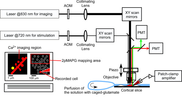
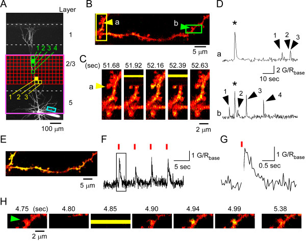
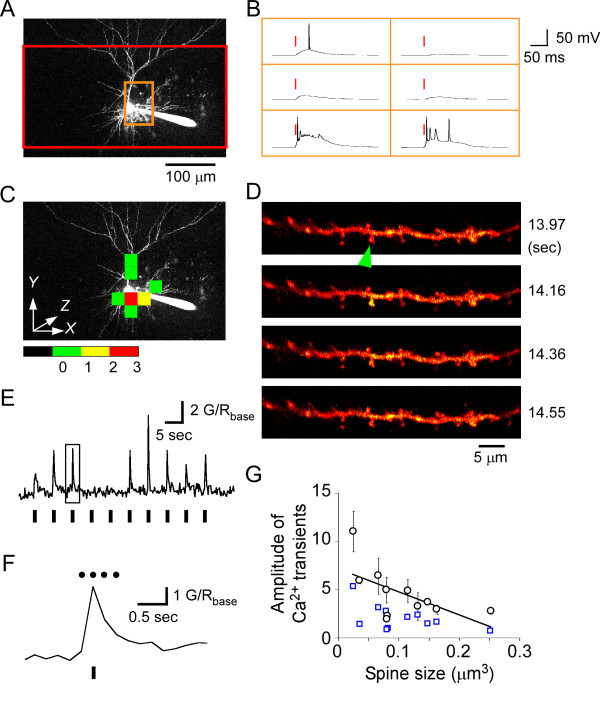
Results: The technique combines two-photon uncaging of glutamate over a broad area (~500 × 250 × 100 μm) with two-photon calcium imaging in a narrow region (~50 × 10 × 1 μm). The former was used for systematic activation of layer 2/3 pyramidal cells in the rat motor cortex, while the latter was used to detect the dendritic spines of layer 5 pyramidal cells that were innervated by some of the photoactivated cells. This technique allowed identification of various sizes of innervated spine located <140 μm laterally from the postsynaptic soma. Spines distal to their parent soma were preferentially innervated by cells on the ipsilateral side. No cluster of neurons innervating the same dendritic branch was detected.
Conclusions: This new method will be a powerful tool for clarifying the microarchitecture of synaptic connections, including the positional and structural characteristics of dendritic spines along the dendrites.

 求助内容:
求助内容: 应助结果提醒方式:


