Scott T Ferry, Hessam M Afshari, Justin A Lee, Laurence E Dahners, Paul S Weinhold
{"title":"Effect of prostaglandin E2 injection on the structural properties of the rat patellar tendon.","authors":"Scott T Ferry, Hessam M Afshari, Justin A Lee, Laurence E Dahners, Paul S Weinhold","doi":"10.1186/1758-2555-4-2","DOIUrl":null,"url":null,"abstract":"<p><strong>Background: </strong>Increased tendon production of the inflammatory mediator prostaglandin E2 (PGE2) has been suggested to be a potential etiologic agent in the development of tendinopathy. Repeated injection of PGE2 into tendon has been proposed as a potential animal model for studying treatments for tendinopathy. In contrast, nonsteroidal anti-inflammatory drugs (NSAIDs) which inhibit PGE2 production and are commonly prescribed in treating tendinopathy have been shown to impair the healing of tendon after acute injury in animal models. The contradictory literature suggests the need to better define the functional effects of PGE2 on tendon. Our objective was to characterize the effects of PGE2 injection on the biomechanical and biochemical properties of tendon and the activity of the animals. Our hypothesis was that weekly PGE2 injection to the rat patellar tendon would lead to inferior biomechanical properties.</p><p><strong>Methods: </strong>Forty rats were divided equally into four groups. Three groups were followed for 4 weeks with the following peritendinous injection procedures: No injection (control), 4 weekly injections of saline (saline), 4 weekly injections of 800 ng PGE2 (PGE2-4 wks). The fourth group received 4 weekly injections of 800 ng PGE2 initially and was followed for a total of 8 weeks. All animals were injected bilaterally. The main outcome measurements included: the structural and material properties of the patellar tendon under tensile loading to failure, tendon collagen content, and weekly animal activity scores.</p><p><strong>Results: </strong>The ultimate load of PGE2-4 wks tendons at 4 weeks was significantly greater than control or saline group tendons. The stiffness and elastic modulus of the PGE2 injected tendons at 8 weeks was significantly greater than the control or saline tendons. No differences in animal activity, collagen content, or mean fibril diameter were observed between groups.</p><p><strong>Conclusions: </strong>Four weekly peritendinous injections of PGE2 to the rat patellar tendon were not found to be an effective model of clinical tendinopathy. In contrast, improved structural and material properties of the patellar tendon were found after PGE2 injection. While PGE2 has been thought to have a contributory role in the development of tendinopathy and anti-inflammatory medications remain a common treatment, our results suggest a positive role of PGE2 in tendon remodeling in some circumstances.</p>","PeriodicalId":88316,"journal":{"name":"Sports medicine, arthroscopy, rehabilitation, therapy & technology : SMARTT","volume":"4 1","pages":"2"},"PeriodicalIF":0.0000,"publicationDate":"2012-01-09","publicationTypes":"Journal Article","fieldsOfStudy":null,"isOpenAccess":false,"openAccessPdf":"https://sci-hub-pdf.com/10.1186/1758-2555-4-2","citationCount":"28","resultStr":null,"platform":"Semanticscholar","paperid":null,"PeriodicalName":"Sports medicine, arthroscopy, rehabilitation, therapy & technology : SMARTT","FirstCategoryId":"1085","ListUrlMain":"https://doi.org/10.1186/1758-2555-4-2","RegionNum":0,"RegionCategory":null,"ArticlePicture":[],"TitleCN":null,"AbstractTextCN":null,"PMCID":null,"EPubDate":"","PubModel":"","JCR":"","JCRName":"","Score":null,"Total":0}
引用次数: 28
Abstract
Background: Increased tendon production of the inflammatory mediator prostaglandin E2 (PGE2) has been suggested to be a potential etiologic agent in the development of tendinopathy. Repeated injection of PGE2 into tendon has been proposed as a potential animal model for studying treatments for tendinopathy. In contrast, nonsteroidal anti-inflammatory drugs (NSAIDs) which inhibit PGE2 production and are commonly prescribed in treating tendinopathy have been shown to impair the healing of tendon after acute injury in animal models. The contradictory literature suggests the need to better define the functional effects of PGE2 on tendon. Our objective was to characterize the effects of PGE2 injection on the biomechanical and biochemical properties of tendon and the activity of the animals. Our hypothesis was that weekly PGE2 injection to the rat patellar tendon would lead to inferior biomechanical properties.
Methods: Forty rats were divided equally into four groups. Three groups were followed for 4 weeks with the following peritendinous injection procedures: No injection (control), 4 weekly injections of saline (saline), 4 weekly injections of 800 ng PGE2 (PGE2-4 wks). The fourth group received 4 weekly injections of 800 ng PGE2 initially and was followed for a total of 8 weeks. All animals were injected bilaterally. The main outcome measurements included: the structural and material properties of the patellar tendon under tensile loading to failure, tendon collagen content, and weekly animal activity scores.
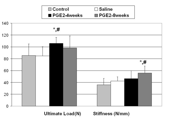
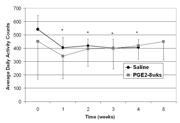
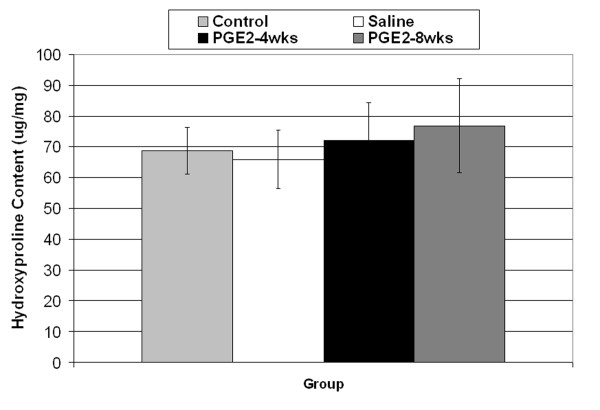
Results: The ultimate load of PGE2-4 wks tendons at 4 weeks was significantly greater than control or saline group tendons. The stiffness and elastic modulus of the PGE2 injected tendons at 8 weeks was significantly greater than the control or saline tendons. No differences in animal activity, collagen content, or mean fibril diameter were observed between groups.
Conclusions: Four weekly peritendinous injections of PGE2 to the rat patellar tendon were not found to be an effective model of clinical tendinopathy. In contrast, improved structural and material properties of the patellar tendon were found after PGE2 injection. While PGE2 has been thought to have a contributory role in the development of tendinopathy and anti-inflammatory medications remain a common treatment, our results suggest a positive role of PGE2 in tendon remodeling in some circumstances.

 求助内容:
求助内容: 应助结果提醒方式:


